MOLECULAR & CELLULAR NEUROBIOLOGY 
Master Course Cognitive Neuroscience - Radboud University, Nijmegen


INDEX

INTRODUCTION CELLS AND WITHIN CELLS IN A NUTSHELL GENOMICS MOLECULAR BIOLOGICAL RESEARCH METHODOLOGY NEURODEVELOPMENT  


Chapter 2:  Cells and within cells


Cells

DNA and genes

Translation

Receptor Mechanisms


    Neurons

   More on DNA

   Proteins, Protein Structure and Protein Analysis

   Ion channel receptors


    Glia

   Epigenetics

   Protein folding in the cell

   Tyrosine kinase receptors

Within cells

   Transcription

   Post-translational modifications of proteins

   G-protein-coupled receptors
   Amino ac, Carbohydr, Lipids and Nucleic ac

   Noncoding RNAs

   Protein degradation in the cell - Autophagy

   G-proteins

   Membranes and Membrane Proteins

   miRNAs and the brain

   Protein secretion / Secretory pathway

   Transcription and signalling

   The Exctracellular Matrix

       Transcription factor receptors


Transcription factor receptors 

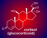
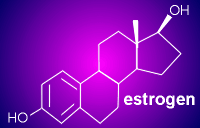
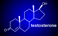
estrogen.gif (4050 bytes) Steroid hormones, thyroid hormones and vitamin A metabolites (called retinoids) regulate the expression of complex gene programs by binding to members of a nuclear receptor family of ligand-activated transcription factors. Illustrated here are the structures of some of the important steroids. The ligands for the transcription factor receptors have the shared property that they are relatively small and lipophillic. Thus, they can easily cross the lipid bilayer of the cell to bind with their intracellular receptor. The receptor for glucocorticoids (cortisol in man, corticosterone in rodents) is often considered the protype of a transcription factor receptor. Therefore, this module will primarily concern the glucocorticoid receptor.

Before considering the glucocorticoids, first use the link to the left to learn more about steroids in general.
 

testosterone.gif (3732 bytes)
butsteroids.gif (5844 bytes) cortisol.gif (4663 bytes)


What are glucocorticoids?  

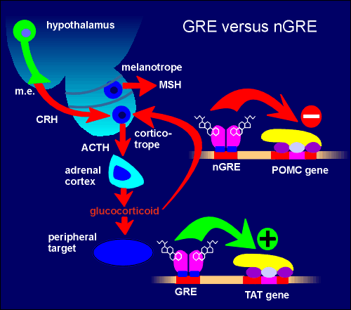
HHAaxis.gif (14198 bytes)Glucocorticoids are steroid hormones synthesized and released by the adrenal cortex. Their general effect in the periphery is a mobilization of glucose (e.g. glycolysis in the liver, gluconeogenesis in other tissues), making glucose available as an energy source. Synthesis and release of glucocorticoids is regulated by the so-called hypothalamo-hypophyseal adrenal axis (fig. to right; hypophyseal = pituitary; so also called hypothalamo-pituitary-adrenal, HPA, axis). Neurons in the hypothalamus of the brain produce the peptide corticotropin-releasing hormone (CRH) which they release to the blood in the median eminence (m.e.). CRH stimulates production and secretion of corticotropin (also called adrenocoroticotropic hormone, ACTH) from corticotrope cells in the pars distalis of the pituitary gland.

bbb.gif (17230 bytes)Being hydrophobic, the glucocorticoids can cross the blood-brain barrier (BBB) and enter brain tissue. This is illustrated in the highly schematic figure to the left where peripheral tissue is indicated in blue and central nervous system tissue in green. By being able to act on both central and peripheral targets the glucocorticoids bring about a coordination of events in these two compartments of the animal. Glucocorticoids give a general activation to peripheral systems and centrally they lead to a general activation of the nervous tissue.


The glucocorticoid receptor is a transcription factor

Similar to all nuclear hormone receptors, the glucocorticoid receptor has a modular structure, consisting of a DNA-binding domain (DBD), a ligand-binding domain and transactivation domains to activate RNA polymerase. One region involved in transactivation is in the N terminal region of the receptor and another region with transactivation function is found within the ligand binding domain of the receptor.The DNA-binding domain is found approximately in the middle of the protein sequence. Here there are zinc fingers with 4 highly conserved cysteine molecules coordinating the binding of the zinc atom. The cysteine amino acids are indicated in yellow in the figure to the left, other amino acids in blue or purple. The formation of zinc fingers results in a structure that interacts with specific DNA sequences within the glucorticoid response element (GRE). Toward the C-terminal is the ligand binding domain. This contains a transactivation domain (already mentioned) and a homodimerization domain responsible for holding the receptor together as a dimer. The ligand binding domain is known to possess two large hyrdrphobic cavities for binding of the glucocorticoid.


The glucocorticoid receptor can activate or repress genes

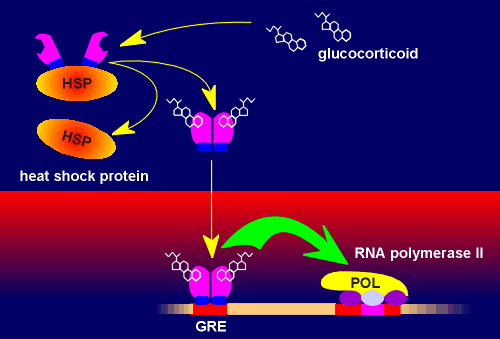
The classical mode of action of the glucocorticoid receptor is via a glucocorticoid response element (GRE) on the DNA. In the absence of ligand, the receptor is trapped as an inactive cytosolic complex together with a family of proteins called heat-shock proteins. Upon ligand binding this complex dissociates and receptor dimers are formed which then translocate to the nucleus. Here it binds to the GRE. The GRE is a highly conserved sequence within the DNA. The bound receptor dimers can now interact with the basal transcriptional machinery (RNA polymerase and associated basal transcription factors). This leads to higher rates of gene transcription.

nGRE.gif (8221 bytes)The glucocorticoid can also act as a DNA binding-dependent repressor. In this case it binds to negative glucocorticoid responsive elements (nGRE) to bring about an inhibition on the basal transcription machinery. The nucleotide sequence in nGRE is less conserved that of GRE but can be identified in a number of genes. In peripheral targets the glucocorticoids signal mostly through GRE.

For example, the gene coding for tyrosine aminotransferase (TAT), one of the important enzymes for gluconeogenesis, has a GRE associated with it.

A good example of a gene that is regulated through nGRE is the proopiomelanocortin (POMC) gene in the corticotropes of the pars distalis of the pituitary gland. In this tissue, POMC is the the precursor protein for the production of adrenocorticotropic hormone (ACTH). Through the nGRE the glucocorticoid brings about an inhibition in the production of ACTH. This is an example of negative feedback and can be regarded as a mechanism to ensure that there is not an overactivation of the hypothalamo-hypophyseal adrenal axis. Interestingly, the action of nGRE on the POMC appears to be tissue specific. For example, glucocorticoids have no effect on POMC gene expression in the melanotrope cell of the pars intermedia where the precursor protein is used to produce the peptide melanophore stimulating hormone (MSH). The ability to act through GREs or nGREs makes glucocoticoid signaling very versatile.

GRclassical.gif (19626 bytes)


negative.gif (25507 bytes)

The glucocorticoid receptor can act through DNA binding-independent mechanisms   


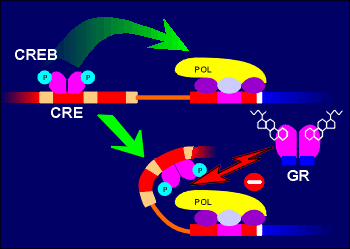
GR CREB.gif (12463 bytes)Adding to the versatility the glucocorticoid receptor in regulatory processes has been the finding that this receptor can also regulate gene expression by influencing the action of other transcription factors, rather than  binding directly to responsive elements on the DNA. These DNA-binding independent mechanisms can be positive or negative. They involve protein-protein interaction between the activated glucocorticoid receptor and other transcription factors or cofactors of transcription factors. For example, the glucocorticoid receptor can bind to  cAMP response element binding protein CREB (possibly through an intermediate factor) and prevent transactivation by CREB. The net result of this action is gene repression.  


The glucorticoid receptor acts in a similar fashion to transcription factors which act on AP-1 sites. In addition it has also been proposed that the glucocorticoid receptor inhibits cJun phosphorylation, a prerequisite for AP-1 activation. Again, the mechanism is DNA binding-independent and the net result is gene repression. Positive or synergistic effects of the glucocorticoid receptor with other transcription factors have also been reported, adding yet more versatility to glucocorticoid receptor signaling.

GR cJun.gif (7672 bytes)

 Dissecting the mechanism of glucocorticoid signaling   

To distinguish DNA binding-dependent and -independent mechanisms of the glucocorticoid receptor an innovative approach has been developed. DBDmutation.gif (14812 bytes)This approach involves introducing a mutation into the DNA-binding domain (DBD) of the glucocorticoid receptor such that it can no longer bind glucorticoid responsive elements on the DNA. Thus, all DNA-dependent mechanisms of the receptor are lost (i.e. direct transactivation or repression via GRE or nGRE). The DNA-independent protein-protein interaction with other transcription factor are left intact. This type of analysis starts by producing a mouse homozygous for the mutated gene (i.e. a DBD-/DBD- mouse). The methods used are the same as those used to produce gene knock-out mice (see "gene transfer").

GRko.gif (5955 bytes)While a glucocorticoid receptor knock-out mouse is lethal (it dies immediately after birth) a mouse homozygous for the mutation in the DBD is viable. This nicely illustrates how important the DNA-independent mechanisms are to glucocorticoid signaling (i.e. with them you survive, without them you die). Glucocorticoid signaling can be examined in these homozygous DBD-/DBD- mice to determine which actions of glucocorticoids are through the DNA binding-dependent and -independent mechanisms. Some interesting divisions of labor between the two mechanisms has been revealed with this approach. As already mentioned, the glucocorticoid receptor inhibits POMC expression via DNA binding-dependent nGRE mechanism. Thus glucocorticoid failed to block POMC expression in DBD-/DBD- mice because the receptor lacked the DNA binding domain. DBD-/DBD- mice had elevated levels of POMC gene expression. Surprisingly, glucocorticoid treatment was found to inhibit ACTH release from the corticotrope cellloops.gif (21704 bytes) in these same animals. This indicates that there are two mechanisms in play for the so-called short loop negative feedback of glucocorticoid on the corticotrope cell: a DNA binding-dependent mechanism for blocking gene expression and a DNA binding-independent mechanism for blocking secretion (perhaps through blocking expression of some protein critical to the exocytosis machinery).

butLOOPS.gif (3242 bytes)Besides short loop feedback, glucocorticoids also display long loop negative feedback on the expression of the gene coding for CRH in hyothalamic neurons (use link to left for more information on feedback loops). In DBD-/DBD- animals glucocorticoid treatment was found to block CRH expression, indicating that this too is via DNA binding-independent mechanisms. There is some evidence that the long loop may be due to the glucocorticoid receptor interfering with CREB signaling on the CRH gene. The analysis of the mechanisms of action of the glucocorticoid receptor in different brain and peripheral targets is of more that just academic interest. There is a clear medical motivation for trying to acquire a better understanding of the molecular mechanisms involved. It will be desirable to develop drugs with are able to distinguish between different modes of action of the glucocorticoid receptors. In this way specific functions of glucocorticoid function can be targeted and deleterious side effects of glucocorticoid therapy might be avoided.


How about other steroid receptors.......   

The receptors for other steroid receptors such as for estrogen, progesterone, tesosterone and aldosterone function very similar to the glucocorticoid receptor. They too can have DNA-dependent and DNA-independent mechanisms. One (minor) difference is that some of these receptors are already in the nucleus in their inactive form (e.g. estrogen receptor) whereas other reside in the cytoplasm in their inactive form.


Next page: From genome to genetic........ Go back to:  Transcription and signalling