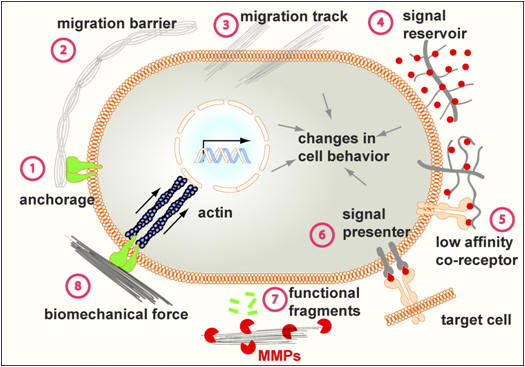
1. Structural proteins: collagen and elastin.
2. Specialized proteins: e.g. fibrillin, fibronectin, and laminin.
3. Proteoglycans: these are composed of a protein core to which is attached long chains of repeating disaccharide units termed glycosaminoglycans (GAGs) forming extremely complex high-molecular weight components of the ECM.
Collagens
Collagens are the most abundant proteins found in the animal kingdom and the major proteins comprising the ECM. There are at least 30 different collagen genes dispersed through the human genome. These 30 genes generate proteins that combine in a variety of ways to create over 20 different types of collagen fibrils in the various ECMs of the body. Collagens are predominantly synthesized by fibroblasts but epithelial cells also synthesize these proteins. The fundamental higher-order structure of collagens is a long and thin diameter rod-like protein. Collagens are synthesized as longer precursor proteins called procollagens. Collagen fibers begin to assemble in the endoplasmic reticulum (ER) and Golgi complexes. The signal sequence is removed and numerous modifications take place in the collagen chains. Alterations in collagen structure resulting from abnormal genes or abnormal processing of collagen proteins results in numerous diseases.
Fibronectins
The role of fibronectins is to attach cells to a variety of extracellular matrices. Fibronectin attaches cells to all matrices except type IV that involves laminin as the adhesive molecule. Fibronectins are dimers of two similar peptides. At least 20 different fibronectin chains have been identified that arise by alternative RNA splicing of the primary transcript from a single fibronectin gene. Fibronectins contain at least six tightly folded domains each with a high affinity for a different substrate such as heparan sulfate, collagen, fibrin and cell-surface receptors. The cell-surface receptor-binding domain contains a consensus amino acid sequence, RGDS.
Lamina
All basal laminae contain a common set of proteins and GAGs: type IV collagen, heparan sulfate proteoglycans, entactin and laminin. The basal lamina is often refered to as the type IV matrix. Each of the components of the basal lamina is synthesized by the cells that rest upon it. Laminin anchors cell surfaces to the basal lamina.
Representative matrix types produced by vertebrate cells
| Collagen | Anchor | Proteoglycan | Cell-Surface Receptor | Cells |
| I | fibronectin | chondroitin and dermatan sulfates | integrin | fibroblasts |
| II | fibronectin | chondroitin sulfate | integrin | chondrocytes |
| III | fibronectin | heparan sulfate and heparin | integrin | quiescent hepatocytes, epithelial; assoc. fibroblasts |
| IV | laminin | heparan sulfate and heparin | laminin receptors | all epithelial cells, endothelial cells, regenerating hepatocytes |
| V | fibronectin | heparan sulfate and heparin | integrin | quiescent fibroblasts |
| VI | fibronectin | heparan sulfate | integrin | quiescent fibroblasts |
