MOLECULAR & CELLULAR NEUROBIOLOGY 
Master Course Cognitive Neuroscience - Radboud University, Nijmegen


INDEX

INTRODUCTION CELLS AND WITHIN CELLS IN A NUTSHELL GENOMICS MOLECULAR BIOLOGICAL RESEARCH METHODOLOGY NEURODEVELOPMENT  


Chapter 2:  Cells and within cells


Cells

DNA and genes

Translation

Receptor Mechanisms


    Neurons

   More on DNA

   Proteins, Protein Structure and Protein Analysis

   Ion channel receptors


    Glia

   Epigenetics

   Protein folding in the cell

   Tyrosine kinase receptors

Within cells

   Transcription

   Post-translational modifications of proteins

   G-protein-coupled receptors
   Amino ac, Carbohydr, Lipids and Nucleic ac

   Noncoding RNAs

   Protein degradation in the cell - Autophagy

   G-proteins

   Membranes and Membrane Proteins

   miRNAs and the brain

   Protein secretion / Secretory pathway

   Transcription and signalling

   The Exctracellular Matrix

       Transcription factor receptors


miRNAs and the brain


miRNAs in the central nervous system (CNS)

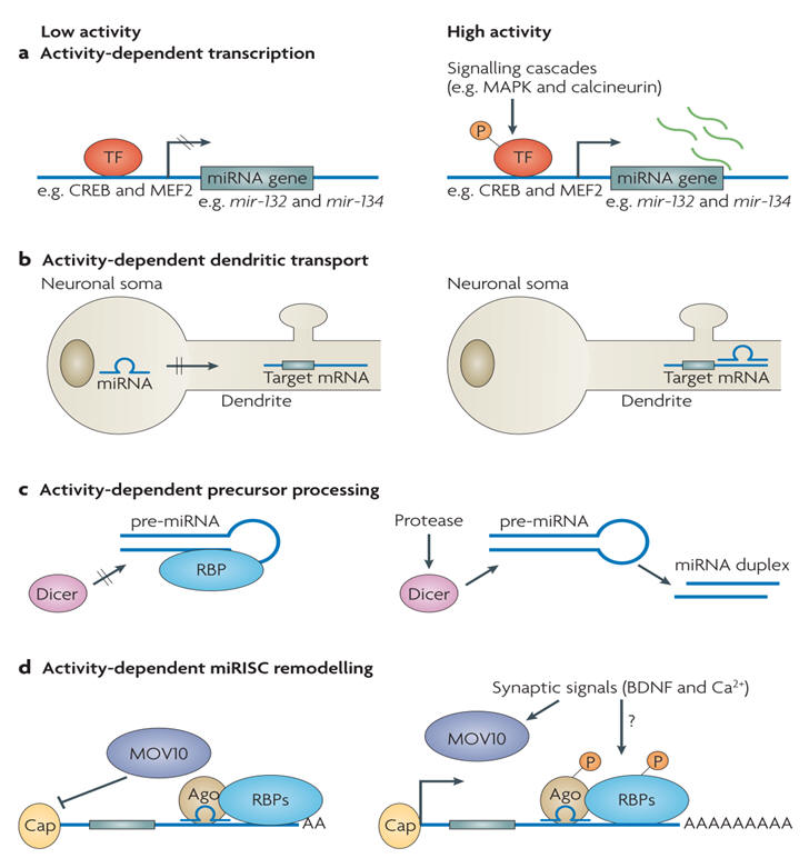
Over half of the miRNAs so far identified are expressed predominantly or exclusively in the brain. The role of miRNAs in brain function is only just emerging. There is accumulating evidence to indicate that miRNAs play a key role in the development of the CNS and play an important role in neuronal differentiation (see below). There is now evidence that miRNAs also play a role in controlling neurotransmitter release.

A major challenge is the prediction of mammalian miRNA targets: thousands of human genes may be miRNA targets. Interactions between miRNA and their targets are often classified as a “switch,” “tuning,” or “neutral”. A “switch” interaction is characterized by a miRNA reducing the target protein activity to a negligible level. This differs from “tuning” interactions, which result in the fine modulation of target protein levels to an optimal range for a given physiological and/or developmental state. “Neutral” targets are usually species-specific interactions that have neither a positive nor negative effect on the cell.  

miRNAs and neurodevelopment

 The human transcriptome shows an amazing complexity with major posttranscriptional control of neuronal development by miRNAs. In the nervous system, critical roles for a number of miRNAs in neuronal development or function have been demonstrated in several model organisms (e.g. Lsy-6 and miR-273 in C. elegans, miR-7 in Drosophila, and miR-134, miR-132, miR-138 and members of the miR-200 family in mammals). In particular, miR-9 and miR-124 are specifically expressed in the mammalian nervous system, and their respective nucleotide sequences are 100% identical among many species. Yet, their expression patterns and mRNA targets are less conserved throughout evolution. As a consequence, these miRNAs exhibit diverse context-dependent functions in different aspects of neuronal development, ranging from early neurogenesis and neuronal differentiation to dendritic morphogenesis and synaptic plasticity (see figure below).

miR-9 and miR-124:

-     -  are the most extensively studied neuronal miRNAs

-     -  are among the most ancient animal miRNAs that show cell-type specific expression

 - -  -  are implicated in multiple stages of neuronal development

-     -  may play key roles in the development of new body plans

-     -  their roles in various aspects of neuronal development in different species will serve as an excellent case study to elucidate the functional conservation and divergence of neuronal miRNAs during evolution

Some other neuronal miRNAs also exhibit context-dependent functions in development. Thus, post-transcriptional regulation of spatial and temporal expression levels of protein-coding genes by miRNAs contributes uniquely to the proper development and evolution of the complex nervous system.

A recurring theme seems to be that one or a few mRNA targets account for the majority of the phenotype in a particular developmental or cellular process. This is likely the case for miRNAs as well. The context-dependent functions of miRNAs in neuronal development or other processes could be explained in part by the variations in transcriptome composition in diverse cell types in different species. The ratio of copy numbers between a specific miRNA and its target may also influence its developmental functions. Thus, it will be useful to systematically identify context-dependent targets of a specific miRNA and to study the endogenous activities of specific miRNAs in their physiological contexts. Overall, conserved neuronal miRNAs may assume novel functions, which, together with newly evolved miRNAs, such as those uniquely expressed in the human brain, may contribute to the evolution of this organ.


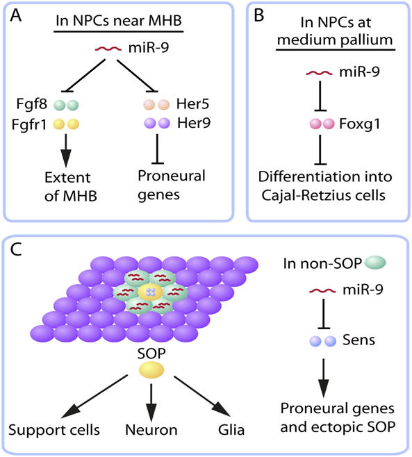
Context-dependent functions of miR-9 in neurogenesis. (A,B) In the developing brains of zebrafish (A) and mice (B), miR-9 is expressed in neural progenitor cells (NPCs) and promotes neurogenesis by downregulating different suppressors of neuronal differentiation. (C) During early neurogenesis in Drosophila embryos, miR-9 is not expressed in sensory organ precursors (SOPs) that eventually give rise to sensory neurons and other cell types. Instead, it is expressed in non-SOP cells, including those adjacent to the SOP in the pro-neural cluster, to suppress the residual expression of Sens, an activator of proneural genes in the process of lateral inhibition. Fgf, fibroblast growth factor; Fgfr, fibroblast growth factor receptor; Foxg1, forkhead box protein G1; MHB, midbrain-hindbrain boundary.


microRNAs at the synapse

Recent evidence points to a widespread role for neural miRNAs at various stages of synaptic development, including dendritogenesis, synapse formation and synapse maturation. Furthermore, studies from invertebrates indicate that miRNAs might contribute to the control of synapse function and plasticity in the adult. Key features of synapse-relevant miRNAs include their ability to regulate mRNA translation locally in the synaptodendritic compartment and the modulation of their expression and function by neuronal activity. The potentially huge impact of miRNA-based mechanisms on higher-order processing, memory and neuropsychiatric disorders in vertebrates is just starting to be recognized.

What features make miRNAs especially well suited to regulate important aspects of synapse development and plasticity? First, a large pool of different miRNA sequences is expressed in post-mitotic neurons at times of synapse development, and many of these miRNAs are associated with translation regulatory complexes. This enormous complexity offers miRNA-regulated pathways the capacity to control the expression of several synapse-relevant proteins simultaneously. In addition, recent estimates indicate that each individual miRNA usually has up to a few hundred different target mRNAs, although not every interaction is necessarily of physiological relevance (“neutral” interactions).

Another feature of interest is that many miRNAs do not act as on–off switches, but rather fine-tune gene expression profiles (“tune” interactions). miRNA-mediated fine-tuning usually occurs in the absence of mRNA degradation, a scenario that fits well with the local regulation of dendritic mRNAs during storage and/or activation at the synapse. Related to this, miRNA-directed suppression of mRNA translation has been shown to be reversible in several cases. This is important, because if miRNAs are to play a part in activity-dependent synapse development and plasticity, the dynamic regulation of miRNA function in response to synaptic stimuli is a prerequisite. Similarly, co-expression of neural miRNAs with their target mRNAs is a common feature, giving rise to the hypothesis that they might participate in feedback mechanisms that connect global transcriptional activation with the control of local dendritic protein synthesis.

The regulation of gene expression programmes by neural activity allows neurons to adapt their connectivity to changes in the environment. Many important synaptic regulators are influenced by neuronal activity, and miRNAs are no exception (see figure below).


miRNA regulation by neuronal activity. Several neuronal miRNAs are subject to regulation by neuronal activity at multiple levels. Whereas activity-dependent miRNA transcription (a) and miRNA-induced silencing complex (miRISC) remodelling (d) are experimentally supported, the regulation of miRNA transport (b) and processing (c) by activity is speculative at this point. (a) The transcription of miRNA genes, such as mir-132 and mir-134, is low under basal conditions (left). High activity triggers the activation of intracellular signalling cascades, which in turn activate sequence-specific transcription factors (TF) (such as CREB and MEF2) that are bound in regulatory regions of the genes. Transcription factor activation involves post-transcriptional modifications such as phosphorylation. (b) In conditions of low activity, miRNA transport (either at the level of the mature or the precursor miRNA (pre-miRNA)) is inhibited, preventing accumulation of the miRNA in the synaptodendritic compartment and interaction with dendritic target mRNAs. High activity promotes dendritic miRNA transport, which could involve the regulation of RNA transport granules. (c) Dicer-mediated processing of pre-miRNAs might be blocked under conditions of low activity, for example by virtue of RNA-binding proteins (RBPs) that prevent Dicer from accessing the stem loop sequence. High activity could modify RBPs in a way that allows Dicer to access the pre-miRNA and proceed with cleavage. (d) Under low-activity conditions, the concerted action of miRISC components (for example, Ago and associated RBPs suppresses efficient protein production. The underlying signalling pathways are unknown. BDNF, brain-derived neurotrophic factor.


The local control of protein synthesis seems to be a preferred site of action of neural miRNAs, although a definitive proof that miRNAs function in a localized manner in the neurons of any organism in vivo is still lacking. Examples of miRNAs operating at the whole-neuron level are beginning to be revealed. Elucidating how local and global control of gene expression exerted by miRNAs is coordinated within neurons is likely to enrich our mechanistic understanding of synaptic plasticity, neuronal homeostasis and synaptic tagging (a process by which synaptic activity evokes a transient synapse-specific change that allows the synapse to capture proteins or mRNAs that are needed for stable long-term potentiation and long-term depression). With the ability of miRNAs to simultaneously fine-tune the expression of hundreds of target genes in response to extracellular cues, it is easy to foresee a great potential for miRNA-based therapeutics for the treatment of neurological disorders of complex genetic origin, such as mood disorders and autism-spectrum diseases. Here, the challenge will be to control potential nonspecific, off-target effects, a phenomenon that is well known from the RNA interference field.

 Understanding miRNA regulation of synaptic connectivity

 Analysis of dendrite and synapse formations has revealed that both miRNA “tuning” and “switching” mechanisms (see above) are involved in regulating proper synaptic connectivity. During neuronal development, increases in dendritic arbor complexity have been shown to be an important determinant of synaptic number, size, and function. Transient depolarization, or exposure to neurotrophins, promotes this dendritic arbor morphogenesis. Recent studies have revealed the importance that neuron-enriched miR-132 and miR-134 have in the activity-regulated rapid response changes of dendritic elaboration.

In addition to being involved in neuritic branch elaboration, miRNAs have been shown recently to play an intricate role in dendritic spine development. It appears that    the dendritic spine specifically uses miRNAs to locally regulate morphology and function through the precise modulation of actin cytoskeletal dynamics, i.e. miRNAs control synapse morphogenesis through regulation of the actin cytoskeleton. Also, miRNAs have been shown to control the physiological architecture of the synapse by modulating the abundance and availability of glutamate receptor (GluR) levels at post-synaptic sites.

miRNAs and neurological disorders

Synapses are increasingly recognized as central structures in the aetiology of a number of neurological disorders, including mental retardation, schizophrenia and neurodegenerative disorders. The knowledge of miRNA function at virtually all steps in synapse development has fuelled research into a potential causative function of miRNAs in such diseases. Most informative in this regard have been genetic mouse models that show certain characteristics of neuropsychiatric diseases. In humans, microdeletions at 22q11, for example, dramatically increase the risk of developing schizophrenia. A mouse model of this has been generated, and these mice have impaired expression of mature miRNAs, including miR-134. The behavioural defects in these mice coincide with abnormal dendrite and spine morphogenesis, further supporting a role for miRNAs in the regulation of neural connectivity. One of the hallmarks of schizophrenia is N-methyl-D-aspartate receptor (NMDAR) hypofunction in the prefrontal cortex. miR-219 has been implicated in the control of NMDAR signalling. The observed down regulation of miR-219 in patients with schizophrenia could be part of a compensatory mechanism to restore NMDA signalling.

Cellular assays have uncovered both miRNA regulatory proteins and miRNA targets that are mutated in neuropsychiatric diseases, including FMR1 (Fragile-X-syndrome), LIMK1 (Willliams syndrome; targeted by miR-134) and MECP2 (Rett syndrome; targeted by miR-132). miRNA-dependent fine-tuning of key disease-associated genes could be an important mechanism to maintain neuronal homeostasis and allow neural circuits to adequately respond to environmental insults.

miRNAs and stress

Chronic psychosocial stress produces many changes at the cellular level mediated by glucocorticoids, including compromising cellular defenses and making them more susceptible to insults (ie. free radicals, seizures, etc), reducing cellular energy stores, and decreasing neurogenesis. miRNAs are hypothesized to play a specialized role in cellular responses to stress. During the cellular stress response, miRNAs have the capacity to change from translation suppressors to activators. Because of this modified activity, miRNAs could provide a pivotal role in mediating cellular adaptation to stress. In particular, miRNAs have been implicated in the way cells respond to oxidative stress, nutrient deprivation and DNA damage. Another area where the interplay between stress and miRNAs has been explored is that of maternal care in early life, which has been shown to prime the brain to be either more or less sensitive to stress and, ultimately, to influence adult ability to adapt to stress. Two potential mechanisms that may underlie this phenomenon are (1) miRNA regulation and (2) epigenetics (see figure below). There is some evidence that miRNAs can modulate glucocorticoids and glucocorticoid receptor function.


miRNAs influence the pathophysiology of psychiatric disorders. Genetic and environmental factors can influence behavior via many possible mechanisms. Modifiable changes in epigenetic or miRNA expression patterns, along with more permanent genetic polymorphisms contribute to varying degrees of resiliency or vulnerability. It is hypothesized that these “predisposing” elements couple with environmental conditions (e.g. early life stress) and lead to either healthy or dysregulated mood that persists throughout an individual’s lifetime.


miRNAs as drugs

Current drugs used to treat the psychotic disorders of schizophrenia and bipolar affective disorder largely target monoamine receptors. Another possibility of developing new antipsychotics is by targeting specific miRNAs. miRNAs offer an exciting potential for developing new antipsychotics, although research in the field is at an early stage. Studies of miRNAs have been far more abundant in schizophrenia than in bipolar affective disorder and the view that non-protein-coding genes have an important regulatory role with implications for the genetic liability to psychosis is gaining greater acceptance. The most promising miRNAs so far identified include miR-181, miR-346 and miR-195 in schizophrenia and miR-34a and miR-144 in bipolar disorder; miR-219 may be involved in both disorders.

Researchers have long struggled to understand the molecular basis of psychiatric disorders (see figure above). Any suggested role that miRNAs may play in mental health is still very much in its infancy of being demonstrated. Further studies are however warranted to determine the precise role that some implicated miRNAs may have in the development, progression and treatment of schizophrenia and what common predicted targets and mechanisms may confer susceptibility.

miRNA modulation has recently been shown to influence circadian rhythm, so regulation of circadian rhythms does not appear to occur only through transcriptional feedback mechanisms, but also via miRNA-mediated translational control. This is particularly relevant to the interplay between miRNAs and mood disorders because circadian rhythm is well known to be dysregulated in individuals with bipolar disorder, and may be an appropriate behavioral phenotype for that disorder.


Next page: Translation Go back to: Noncoding RNAs