MOLECULAR & CELLULAR NEUROBIOLOGY 
Master Course Cognitive Neuroscience - Radboud University, Nijmegen


INDEX

INTRODUCTION CELLS AND WITHIN CELLS IN A NUTSHELL GENOMICS MOLECULAR BIOLOGICAL RESEARCH METHODOLOGY NEURODEVELOPMENT  


Chapter 2:  Cells and within cells


Cells

DNA and genes

Translation

Receptor Mechanisms


    Neurons

   More on DNA

   Proteins, Protein Structure and Protein Analysis

   Ion channel receptors


    Glia

   Epigenetics

   Protein folding in the cell

   Tyrosine kinase receptors

Within cells

   Transcription

   Post-translational modifications of proteins

   G-protein-coupled receptors
   Amino ac, Carbohydr, Lipids and Nucleic ac

   Noncoding RNAs

   Protein degradation in the cell - Autophagy

   G-proteins

   Membranes and Membrane Proteins

   miRNAs and the brain

   Protein secretion / Secretory pathway

   Transcription and signalling

   The Exctracellular Matrix

       Transcription factor receptors


Translation: RNA → Protein

As mentioned under "Transcription", gene expression occurs in two steps:

  • transcription of the information encoded in DNA into a molecule of RNA (see under "Transcription") and

  • translation of the information encoded in the nucleotides of mRNA into a defined sequence of amino acids in a protein (i.e. RNA-directed synthesis of polypeptides, described here).

In eukaryotes, the processes of transcription and translation are separated both spatially and in time. Transcription of DNA into mRNA occurs in the nucleus. Translation of mRNA into polypeptides occurs on polysomes in the cytoplasm. In prokaryotes (which have no nucleus), both of these steps of gene expression occur simultaneously: the nascent mRNA molecule begins to be translated even before its transcription from DNA is complete. Although the chemistry of peptide bond formation is relatively simple, the processes leading to the ability to form a peptide bond are exceedingly complex. Translation requires all three classes of RNA. The template for correct addition of individual amino acids is the mRNA, yet both transfer RNAs (tRNAs) and ribosomal RNAs (rRNAs) are involved in the process.

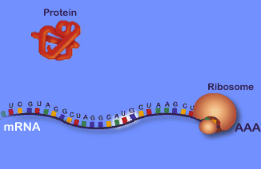
How does a particular sequence of nucleotides specify a particular sequence of amino acids? The answer: by means of  the tRNA molecules, each specific for one amino acid and for a particular triplet of nucleotides in mRNA called a codon. The family of tRNA molecules enables the codons in an mRNA molecule to be translated into the sequence of amino acids in the protein. Translation is thus the part of protein synthesis where the ribosomes in the cytoplasm use tRNA to attach to the mRNA and translate the bases into amino acids and tRNA molecules bring the specified amino acids that the ribosome links together to make a protein.Thus, the tRNAs carry activated amino acids into the ribosome which is composed of rRNA and ribosomal proteins. The ribosome is associated with the mRNA ensuring correct access of activated tRNAs and containing the necessary enzymatic activities to catalyze peptide bond formation.

Protein translation thus occurs when a single mRNA moves along the ribosome and is read by tRNA molecules, which bring the amino acid to the chain. Once a polypeptide is formed from the specific mRNA, it may in itself form the protein or combine with other polypeptides to form the mature protein. Once the protein is formed, it has certain amino acid sequences which direct it to its specific compartment in the cell (see below for a movie on protein transport into the mitochondrion). Secreted protein molecules, for example, contain a hydrophobic tail sequence which directs it to the endoplasmic reticulum membrane. The proteins perform all of the work of the cell and are each synthesized from a unique mRNA. Gene expression refers to the whole process from the formation of the gene to a mature protein. Many proteins undergo chemical modification posttranslationnally, such as glycosylation. This form of posttranslational modification is particularly common with cellular secretory or membrane proteins. It may involve the development of disulfide bonds, proteolytic cleavage of the newly synthesized protein, or the addition of carbohydrate moieties. These posttranslational changes may importantly affect function and subcellular localization of proteins.

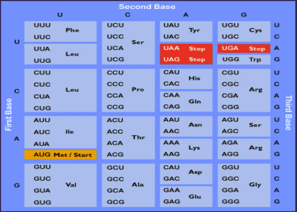
In sum, the genetic code is read in a sequential manner starting near the 5' end of the mRNA. This means that translation proceeds along the mRNA in the 5' ——> 3' direction which corresponds to the N-terminal to C-terminal direction of the amino acid sequences within proteins. The code is composed of a triplet of nucleotides. All 64 possible combinations of the four nucleotides code for amino acids, i.e. the code is degenerate since there are only 20 amino acids. The precise dictionary of the genetic code was determined with the use of in vitro translation systems and polyribonucleotides. The results of these experiments confirmed that some amino acids are encoded by more than one triplet codon, hence the degeneracy of the genetic code. These experiments also established the identity of translational termination codons.

Click here for a movie on "Translation".

Click here for a movie on "Protein Transport".


The steps of translation   

1. Initiation

  • The small subunit of the ribosome (shown in blue) binds to a site "upstream" (on the 5' side) of the start of the message.

  • It proceeds downstream (5' -> 3') until it encounters the start codon AUG. (The region between the cap and the AUG is known as the 5'-untranslated region [5'-UTR].)

  • Here it is joined by the large subunit and a special initiator tRNA; in eukaryotes, initiator tRNA carries methionine (Met).

2. Elongation

  • An aminoacyl-tRNA (a tRNA covalently bound to its amino acid, in this case alanine, Ala) able to base pair with the next codon on the mRNA arrives at the A site (green).

  • The preceding amino acid (Met at the start of translation) is covalently linked to the incoming amino acid with a peptide bond (shown in red).

  • The initiator tRNA is released from the P site.

  • The ribosome moves one codon downstream.

  • This shifts the more recently-arrived tRNA, with its attached peptide, to the P site and opens the A site for the arrival of a new aminoacyl-tRNA.

Note: the initiator tRNA is the only member of the tRNA family that can bind directly to the P site. The P site is so-named because, with the exception of initiator tRNA, it binds only to a peptidyl-tRNA molecule; that is, a tRNA with the growing peptide attached.The A site is so-named because it binds only to the incoming aminoacyl-tRNA; that is the tRNA bringing the next amino acid. So, for example, the tRNA that brings Met into the interior of the polypeptide can bind only to the A site.


3. Termination

  • The end of translation occurs when the ribosome reaches one or more STOP codons (UAA, UAG, UGA). (The nucleotides from this point to the poly(A) tail make up the 3'-untranslated region [3'-UTR] of the mRNA.)

  • There are no tRNA molecules with anticodons for STOP codons.

  • However, a protein release factor recognizes these codons when they arrive at the A site; binding of this protein releases the polypeptide from the ribosome.

  • The ribosome splits into its subunits, which can later be reassembled for another round of protein synthesis.

The Genetic Code - The RNA Codons

Shown below are the triplets that are used for each of the 20 amino acids found in eukaryotic proteins. The row on the left side indicates the first nucleotide of each triplet and the row across the top represents the second nucleotide. The wobble position nucleotides are indicated in blue. The three stop codons are highlighted in red.

                The codon triplets of the genetic code                  

Note:

  • Most of the amino acids are encoded by synonymous codons that differ in the third position of the codon.

  • In some cases, a single tRNA can recognize two or more of these synonymous codons.

  • Example: phenylalanine tRNA with the anticodon 3' AAG 5' recognizes not only UUC but also UUU.

  • The violation of the usual rules of base pairing at the third nucleotide of a codon is called "wobble"

  • The codon AUG serves two related functions

    • It begins every message; that is, it signals the start of translation placing the amino acid methionine at the amino terminal of the polypeptide to be synthesized.

    • When it occurs within a message, it guides the incorporation of methionine.

  • Three codons, UAA, UAG, and UGA, act as signals to terminate translation. They are called STOP codons.

Protein Synthesis Inhibitors

Many of the antibiotics utilized for the treatment of bacterial infections as well as certain toxins function through the inhibition of translation. Inhibition can be effected at all stages of translation from initiation to elongation to termination.

Several Antibiotic and Toxin inhibitors of Translation

Inhibitor

Comments

Chloramphenicol

inhibits prokaryotic peptidyl transferase

Structure of chloramphenicol

Streptomycin

inhibits prokaryotic peptide chain initiation, also induces mRNA misreading

Tetracycline

inhibits prokaryotic aminoacyl-tRNA binding to the ribosome small subunit

Structure of tetracycline

Neomycin

similar in activity to streptomycin

Erythromycin

inhibits prokaryotic translocation through the ribosome large subunit

Fusidic acid

similar to erythromycin only by preventing EFG from dissociating from the large subunit

Puromycin

resembles an aminoacyl-tRNA, interferes with peptide transfer resulting in premature termination in both prokaryotes and eukaryotes

Diphtheria (diptheria) toxin

protein from Corynebacterium diphtheriae which which causes diphtheria (diptheria); catalyzes ADP-ribosylation and inactivation of eEF-2; eEF-2 contains a modified His residue known as diphthamide (dipthamide), it is this resudue that is the target of the toxin

Structure of diphthamide (dipthamide) residueADP-ribosylated diphthamide (dipthamide) residue

Ricin

found in castor beans, catalyzes cleavage of the eukaryotic large subunit rRNA

Cycloheximide

inhibits eukaryotic peptidyltransferase

Structure of cycloheximide


Next page: Proteins, Protein Structure and Protein Analysis Go back to:   miRNAs and the brain