MOLECULAR & CELLULAR NEUROBIOLOGY 
Master Course Cognitive Neuroscience - Radboud University, Nijmegen


INDEX

INTRODUCTION CELLS AND WITHIN CELLS IN A NUTSHELL GENOMICS MOLECULAR BIOLOGICAL RESEARCH METHODOLOGY NEURODEVELOPMENT  


Chapter 2:  Cells and within cells


Cells

DNA and genes

Translation

Receptor Mechanisms


    Neurons

   More on DNA

   Proteins, Protein Structure and Protein Analysis

   Ion channel receptors


    Glia

   Epigenetics

   Protein folding in the cell

   Tyrosine kinase receptors

Within cells

   Transcription

   Post-translational modifications of proteins

   G-protein-coupled receptors
   Amino ac, Carbohydr, Lipids and Nucleic ac

   Noncoding RNAs

   Protein degradation in the cell - Autophagy

   G-proteins

   Membranes and Membrane Proteins

   miRNAs and the brain

   Protein secretion / Secretory pathway

   Transcription and signalling

   The Exctracellular Matrix

       Transcription factor receptors


The G-protein-coupled receptors

There are three components which must be considered in receptor signaling through G-proteins. There is

  • (1) the receptor,

  • (2) the G-protein and

  • (3) the effector.

The ligand receptor complex activates the G-protein which in turn activates (or in some cases inactivates) the effector protein. The effector proteins can be enzymes or ion channels. This section will consider the structure and function of the receptors which link to G-proteins. The next part will take a look at the G-proteins in more detail.

To give you an idea of the relatively important position G-protein-coupled receptors (GPCRs) have in cell signaling, particularly with respect to the neuropeptides, the table to the right is included which gives a list of cloned receptors, compiled in 1995. As you can see, classical neurotransmitters very often have both ligand-gated ion channels as receptors and GPCRs. The neuropeptides, without exception (thus far), function through GPCRs.

The G-protein family of receptors is the most rapidly growing family, far outnumbering both the ligand-gated ion channel family of receptors and the tyrosine kinase receptor family. At present  there are at least 300 known GPCRs (some of these are orphan receptors where the ligand is, as yet, unknown). It has been estimated (from the rate of their discovery in the human genome project) that there will ultimately be 1000 to 3000 such receptors (most of them representing taste/smell receptors). Thus, this receptor mechanism has an extremely important place in cell signaling in general and in brain function in particular.

LISTreceptors.gif (15812 bytes)

 Functional domains of the G-protein-coupled receptors

The GPCRs are very large monomeric proteins. They are intimately associated with the membrane, in contrast to ion channel receptors and tyrosine kinase receptors which have very large extracellular regions. The ligand binding in many GPCRs occurs deep in the transmembrane regions of the receptors.


receptors.gif (17638 bytes)

Reflecting the intimate association with the membrane, the GPCRs have 7 transmembrane regions. These transmembrane regions (numbered I-VII) were first discovered in hyropathy profiles when cDNA recombinant methods were applied to this class of receptors. Each transmembrane region consists of an alpha helix of 20 to 25 amino acids. The N-terminal region of GPCRs is extracellular and the C-terminal region is intracellular.


7trans.gif (20405 bytes)

The 7 transmembrane regions form a circle (illustrated to the right). In the search for functional domains of GPCRs considerable efforts have been made to identify ligand binding domains and the domains responsible for binding to and activation of G-proteins. The general approach in research on the GPCRs has been the same as with the ion channel receptors and tyrosine kinase receptors, namely:

    (1) prepare a hydropathy profile from the sequencing information obtained from the cDNA,

    (2) look for potential functional domains within the profile (e.g. ligand binding domain),

    (3) test the potential domains to construct a model of the receptor.


GPRECEP.gif (50161 bytes)

Given to the left is the "streached-out" model of the beta-adrenergic receptor, which is often considered the "prototype" of a GPCR. The beta-adrenergic receptor was the first GPCR to be fully sequenced by the recombinant cDNA approach. The N terminal is extracellular and there is, within this extracellular region, carbohydrate side chains attached to the structure (one is shown). This is termed "glycosylation". The function of the glycosylations is unknown, but it is thought that it may be important for proper folding of the receptor. The 7 alpha-helix transmembrane regions are mostly composed of hydrophobic amino acids (hydrophobic amino acids are indicated with red circles in the model). The above model shows four functional, or potentially functional, domains for the beta adrenergic receptor (ligand-binding domain, switch domain, G-protein binding domains and regulatory domains). These are discussed individually below.

 The ligand-binding domain

butsearch.gif (9847 bytes)

Ligands for most GPCRs bind deep within the transmembrane regions of the receptor (ligand is illustrated in yellow in figure to right). For the beta-adrenergic receptor the amino acids involved in ligand binding have been identified. These are indicated in the  model given above (Asp, indicated in green at position 113 in TM III, Serines indicated in blue at 204 and 207 in TM V and Phe indicated in purple at 209 in TM VI).

A series of experiments using site-directed mutagenesis were conducted to identify the ligand binding sites. Use the link to the left  to obtain more information on how this approach illuminated the  ligand binding domain of the beta adrenergic receptor.


7transligand.gif (22053 bytes)

The switch?

Within the transmembrane regions there are a number of prolines which, as mentioned in the discussion of ion channel receptors, is an unusual finding in a transmembrane region. As with the ion-channel receptors, one idea is that the prolines (through cis-trans conformational changes) would be involved in the transduction of ligand binding into a conformational change of the receptor. In this case the conformational change would alter the structure of the receptor, so it could interact with G-proteins (i.e. induce a change in the structure of G-protein binding domains so they could bind to the G-proteins). However, the idea of a switch function for the prolines has never been rigorously tested.

The G-protein-binding domains

The obvious place to look for the G-protein-binding domains was intracellular and, indeed, the regions responsible for G protein binding has been shown to be the three intracellular loops (see model above). Of the three loops, the 3rd intracellular loop has proved to be the most important in G-protein binding and activation. One important approach in searching for the G-protein-coupling domains has been to introduce mutations in the cDNA (point mutations or eliminating entire segments of the receptor).This mutated DNA is then used as a template to produce RNA for injection into an expression system, such as Xenopus oocytes (see also "gene transfer").


chemera.gif (49881 bytes)

Following translation of the RNA into protein the mutated receptor was examined for its ability to bind G-proteins. Also, receptor chimeras were produced for analysis of G-protein coupling. For example, the cDNA coding for the 3rd intracellular loop of receptor A was spliced into the cDNA coding for receptor B (which had its own 3rd intracellular loop DNA removed). The resulting cDNA coded for a receptor chimera i.e. a receptor from gentically different sources (see figure to right). When the cDNA for this receptor was transcribed to RNA, and the RNA brought to expresssion, the G-protein preference for the receptor chimera could then be analyzed. When this type of experiment was conducted with the beta adrenergic receptor it was found that substitution of the 3rd intracellular loop dramatically impaired its ability to couple to its normal G-protein, a G-protein known as Gs. These types of experiments have demonstrated the importance of the intracellular loops, particularly the 3rd loop, in G-protein binding.

The regulatory domains

regdomain.gif (23368 bytes)


Sequences corresponding to sites for serine / tryrosine kinase phosphorylations have been found in the C-terminal segment and in the third intracellular loop of the beta adrenergic receptor (illustration to left and green boxes in model above). Indeed, the beta adrenergic receptor (and other GPCRs) have been shown to be among the target proteins for kinases. In some cases this forms autoregulatory negative feedback loops and in other cases sites for "cross-talk" between the GPCR mechanism. Thus, just as GPCRs can cross-talk with ion-channel receptors, so too can the GPCRs, via the production of second messengers and activation of kinases, cross-talk with each other.

There are three major classes of G-protein-coupled receptors  

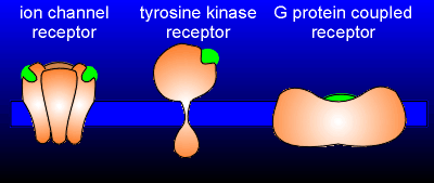
The beta-adrenergic receptor may be considered the prototype for GPCRs, but in fact there are six different classes of receptors in the superfamily of GPCRs. The classes have been constructed on the basis of sequence homology and structure. For the neurotransmitters and neuropeptides, only the first three classes, termed classes A, B and C,  are important.
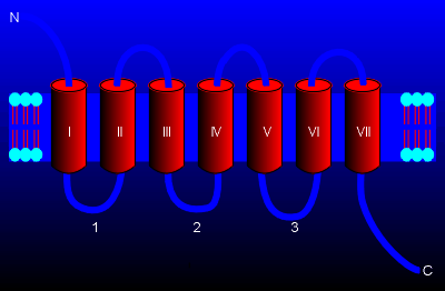
The classes of proteins in the 7 transmembrane (7TM) GPCR superfamily are also often referred to by the name of a prominent member of the class. Thus, we have the rhodopsin-like family, the calcitonin receptor-like family or the metabotropic glutamate receptor family. The 
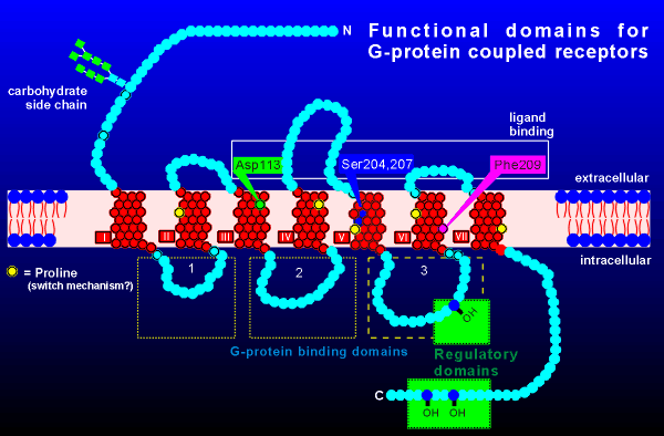
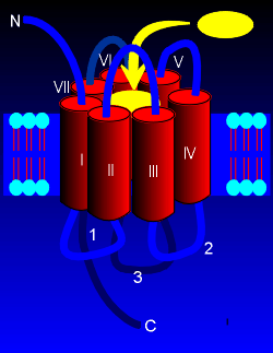
class A receptors include receptors for biogenic amines such as adrenaline, noradrenaline and dopamine. Some of the neuropeptide receptors also come from this family. This class of receptors is characterized by being heavily glycosylated at the N-terminal and possessing a palmitolyation in the intracellular C-terminal region. Palmitoylation concerns the attachment of a lipid group to the amino acid cysteine, the consequence of which is that the lipid plugs into the membrane, thus creating a fourth intracellular loop.

The agonists for class A are usually quite small and their binding sites are often deep in the transmembrane region (e.g. binding of noradrenaline to the beta-adrenergic receptor). Most peptide hormones and neuropeptides have receptors from the Class B family of receptors. Class B receptors have a larger N-terminal extracellular region than class A receptors, and this region possess many disulphide bridges. These bridges may be important in the formation of the globular ligand-binding domain. The ligands for class B receptors can be quite large, such as large proteins. It is thought that upon binding to the extracellular domain the ligand is then in the proper orientation so that another part of the ligand can induce receptor activation through interactions with the transmembrane regions of the receptor. Class C is a very small family, consisting of receptors for only three ligands, namely:

(1) GABA, with the receptor referred to as the GABAb receptor to distinguish it from the ionotropic GABAa receptor,

(2) glutamate, called the metabotropic glutamate receptor to distinguish it from the ionotropic receptors for glutamate, and

(3) calcium, called the Ca2+ sensing receptor, which is important in the regulation of release of hormone involved in Ca2+ homeostasis.

Class C receptors are characterized by very large N-terminal extracellular domains. There are many disulphide bridges in this extracellular region that are important in forming the ligand binding domain. Ligand binding to Class C receptors is believed to induce receptor dimerization which initiates the process of transmembrane signaling.

Interesting points: The sequence homology between the classes of GRPCs is very low. This has led some to suggest that the 7-transmembrane receptors have been reinvented a number of times during evolution. The class E cAMP receptor has only been found in the slime mold Dictyostelium.


Next page: G-proteins Go back to:  Tyrosine kinase receptors