MOLECULAR & CELLULAR NEUROBIOLOGY 
Master Course Cognitive Neuroscience - Radboud University, Nijmegen


INDEX

INTRODUCTION CELLS AND WITHIN CELLS IN A NUTSHELL GENOMICS MOLECULAR BIOLOGICAL RESEARCH METHODOLOGY NEURODEVELOPMENT  


Chapter 2:  Cells and within cells


Cells

DNA and genes

Translation

Receptor Mechanisms


    Neurons

   More on DNA

   Proteins, Protein Structure and Protein Analysis

   Ion channel receptors


    Glia

   Epigenetics

   Protein folding in the cell

   Tyrosine kinase receptors

Within cells

   Transcription

   Post-translational modifications of proteins

   G-protein-coupled receptors
   Amino ac, Carbohydr, Lipids and Nucleic ac

   Noncoding RNAs

   Protein degradation in the cell - Autophagy

   G-proteins

   Membranes and Membrane Proteins

   miRNAs and the brain

   Protein secretion / Secretory pathway

   Transcription and signalling

   The Exctracellular Matrix

       Transcription factor receptors


Protein degradation in the cell

Proteins are continuously synthesised and degraded in all living organisms. The concentration of individual cellular proteins is determined by a balance between the rates of synthesis and degradation, which in turn are controlled by a series of regulated biochemical mechanisms. Differences in the rates of protein synthesis and breakdown result in cellular and tissue atrophy (loss of proteins from cells) and hypertrophy (increase in protein content of cells). The proper functioning of the cell thus requires careful control of the levels of important structural proteins, enzymes and regulatory proteins. The only way that cells can reduce the steady state level of a particular protein is by proteolytic degradation. Thus, complex and highly-regulated mechanisms have been evolved to accomplish this degradation. The degradation rates of proteins are important in determining their cellular concentrations. Proteins break down at rates ranging from 100% per hour to less than 10% per hour and their half-lives (time taken for loss of half the protein molecules) vary between 24 h and 72 h. Regulatory enzymes and regulatory proteins have much shorter half-lives of the order of 5-120 min. Protein breakdown can take place in the mitochondria, chloroplasts, the lumen of the endoplasmic reticulum (ER) and the endosomes, but occurs most commonly in one of two major sites of intracellular proteolysis: lysosomes and the cytosol. The individual degradation rates of proteins vary within a single organelle or cell compartment and also from compartment to compartment, due either to differing sensitivity to local proteases or differing rates of transfer to the cytosol or lysosomes. The range of protein degradation rates within a single organelle is limited, suggesting that the proteins may be treated as groups or families.

Short-lived regulatory proteins are degraded in the cytosol by local proteolytic mechanisms. All short-lived proteins are thought to contain recognition signals that mark them for early degradation. One commonly employed method is the selective labelling of targeted proteins by ubiquitin molecules (see also below). Ubiquitin, a protein of 76 amino acids, binds covalently to available lysine residues on target proteins, which are then recognised by proteases. A number of molecular recognition signals for intracellular protein degradation have been identified, and there are likely to be others as yet undiscovered. Additional degradative mechanisms exist for the identification and rapid degradation of proteins that contain translational or post-translational errors, or have been damaged in some way.

An additional role of intracellular proteolysis is in the stress-response. Cells that are subject to stress, such as starvation, heat-shock, chemical insult or mutation, respond by increasing the rates of proteolysis. One function of this enhanced proteolysis is to salvage amino acids from non-essential proteins. These amino acids can then be reutilized in the synthesis of essential proteins or metabolized directly to provide energy. Another function is in the repair of damage caused by the stress. For example, oxidative stress has been shown to damage a variety of proteins and cause them to be rapidly degraded. Some proteins are genetically unstable. There appear to be signal sequences which target proteins to degradation. These can be part of the protein structure, or added post-translationally.

The two major systems of protein degradation in the cell are the lysosomal pathway and the proteasomal pathway (including ER-Associated Protein Degradation, ERAD; see below).

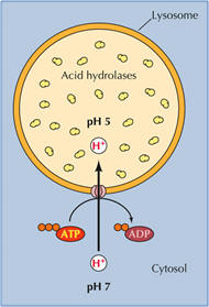
Lysosomal degradation. Most non-selective protein degradation takes place in the lysosomes, where changes in the supply of nutrients and growth factors can influence the rates of protein breakdown. Lysosomes contain a large variety of hydrolytic enzymes that degrade proteins and other substances taken in by endocytosis. Lysosomes have a low internal pH due to activity of vacuolar proton ATPase (see figure). All lysosomal hydrolases exhibit acidic pH optima.

Lysosomal proteases include many cathepsins (cysteine proteases), as well as some aspartate proteases and one zinc protease. Activation may be catalyzed by other lysosomal enzymes or be autocatalytic, promoted by the acidic pH within the lysosome.The rates of lysosomal degradation can vary greatly with cell type and conditions, ranging from less than 1% of total cell protein per hour to 5-10% per hour. The lysosomal degradation of some cytosolic proteins increases in cells deprived of nutrients. It is assumed that the proteins undergoing enhanced degradation are of limited importance for cell viability, and can be sacrificed to support the continuing synthesis of key proteins.

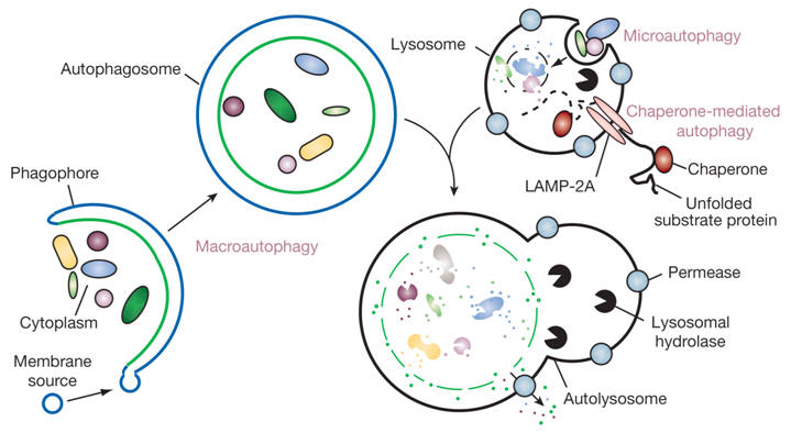
Proteins enter lysosomes by macroautophagy, that is the enclosure of a volume of the cytoplasm by an intracellular membrane. In autophagy, part of the cytoplasm may become surrounded by two concentric membranes. Fusion of the outer membrane of this autophagosome with a lysosomal vesicle results in degradation of enclosed cytoplasmic structures and macromolecules. Autophagy is not a mechanism for selective degradation of individual macromolecules.


Recognizing and destroying proteins that are unable to fold in the ER   

A major function of the ER chaperones is to promote protein folding by preventing misfolding or aggregation. During conditions of ER stress, alterations in the ER environment can profoundly affect the folding of many proteins. Although BiP appears to be the sole chaperone that is monitored by the cell to sense ER stress, many of the chaperones are coordinately up-regulated. The main function of the increased levels of ER chaperones is to bind to unfolded proteins, prevent them from aggregating, and to aid and monitor their refolding if normal physiological conditions are restored to the ER. ER chaperones thus contribute to or control all of the major functions of the ER, including translocation of nascent polypeptide chains, folding and assembly of secretory pathway proteins, monitoring the success of this operation, identifying those proteins that fail this quality control and targeting them for degradation, and finally a role in storing calcium in the ER, which is important in cellular signal transduction pathways, and which also may play a role in initiating apoptosis when ER stress is sustained. Proteins that have ultimately failed ER quality control are degraded to prevent their accumulation in the ER. Accumulation might either titrate out the components of the chaperone systems or form large insoluble aggregates that would be toxic to the cell. The turnover mechanism is termed ERAD, which is conserved from lower eukaryotes like yeast to mammals. ERAD is a process by which misfolded ER proteins are detected and prevented from progressing along the secretory pathway, and directed to the translocon for retrotranslocation (or dislocation) into the cytosol, where they undergo ubiquitin- and proteasome-dependent degradation (cytoplasmic ubiquitin-proteasome system, UPS). Ubiquitin (Ub) is a highly conserved small protein that is universally expressed in eukaryotic cells. Ubiquitination of substrates is a multi-step process. Thus, a link exists between the turnover of ER membrane proteins and UPS. Retrotranslocation may utilize the same core protein complex that forms the protein conducting channel in the translocon through which proteins are delivered to the ER lumen. ERAD thus eliminates misfolded or unassembled proteins from the ER. ERAD targets are selected by a quality control system within the ER lumen (calnexin and BiP play sequential roles in identifying and targeting ERAD substrates for degradation) and are ultimately destroyed by the UPS.


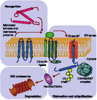
Proteasomal degradation of ERAD targets. Aberrant proteins are recognized within the ER lumen by different quality control mechanisms, which escort terminally misfolded polypeptides to a putative channel that facilitates their export from the ER. Cytoplasmically exposed lysine residues are ubiquitinated by ubiquitin ligases. Dislocation is completed with the help of a protein complex (Cdc48p/p97) and membrane-extracted substrates are conveyed to the proteasome by accessory factors (such as Rad23p and Dsk2p).


The cell's waste disposer, the proteasome. The black spots indicate active, protein-degrading surfaces.


Ubiquitin-mediated protein degradation

  1. The E1 enzyme activates the ubiquitin molecule. This reaction requires energy in the form of ATP.
  2. The ubiquitin molecule is transferred to a different enzyme, E2.
  3. The E3 enzyme can recognise the protein target which is to be destroyed. The E2-ubiquitin complex binds so near to the protein target that the actual ubiquitin label can be transferred from E2 to the target.
  4. The E3 enzyme now releases the ubiquitin-labelled protein.
  5. This last step is repeated until the protein has a short chain of ubiquitin molecules attached to itself.
This ubiquitin chain is recognised in the opening of the proteasome. The ubiquitin label is disconnected and the protein is admitted and chopped into small pieces.


ERAD and diseases      

Because ERAD is a central element of the secretory pathway, it is not surprising that it has major implications for the generation of human diseases. The underlying disorders can be classified into two groups. The first group results from loss-of-function mutations in ERAD components that stabilize aberrant proteins, which in turn accumulate and damage the cell. A prominent example of this is Parkinson's disease. Many cases of familial Parkinson's disease are associated with mutations in the Parkin gene linking ERAD to the generation of this neurodegenerative disorder. The Parkin protein functions as a ubiquitin ligase. The second group of disorders is caused by premature degradation of secretory or membrane proteins, preventing their deployment to distal compartments, as is the case in cystic fibrosis. Bypassing ERAD is not a viable therapeutic strategy because it could yield elevated levels of other ER proteins which could be harmful.

See also under "Protein folding in the cell".


Autophagy   

Until recently, autophagy or cellular self-digestion (a cellular pathway involved in protein and organelle degradation), was thought of primarily as part of the cell's garbage disposal system. Now it is known to be involved in cellular protein and organelle degradation during development as well as during adaptations to changing environmental conditions. Furthermore, autophagy has  an astonishing number of connections to human disease. For example, autophagic dysfunction is associated with cancer, neurodegeneration, microbial infection and ageing. Paradoxically, although autophagy is primarily a protective process for the cell, it can also play a role in cell death. Understanding autophagy may ultimately allow scientists and clinicians to harness this process for the purpose of improving human health.

 At first glance, it may seem perplexing that a process of cellular self-eating could be beneficial. In its simplest form, however, autophagy probably represents a single cell's adaptation to starvation—if there is no food available in the surroundings, a cell is forced to break down part of its own reserves to stay alive until the situation improves. In single-cell organisms such as yeasts, this starvation response is one of the primary functions of autophagy, but in fact this role extends up through to humans. For example, even on a day-to-day basis, autophagy is activated between meals in organs such as the liver to maintain its metabolic functions, supplying amino acids and energy through catabolism.

 There are various types of autophagy, including micro- and macroautophagy, as well as chaperone-mediated autophagy (CMA), and they differ in their mechanisms and functions (Figure 1).  Both micro- and macroautophagy have the capacity to engulf large structures through both selective and non-selective mechanisms, whereas CMA degrades only soluble proteins, albeit in a selective manner. The capacity for large-scale degradation is important in autophagic function, but it carries a certain risk, because unregulated degradation of the cytoplasm is likely to be lethal. On the other hand, basal levels of autophagy are important for maintaining normal cellular homeostasis. Thus, it is important that autophagy be tightly regulated so that it is induced when needed, but otherwise maintained at a basal level.


Figure 1. Different types of autophagy. Microautophagy refers to the sequestration of cytosolic components directly by lysosomes through invaginations in their limiting membrane. The function of this process in higher eukaryotes is not known, whereas microautophagy-like processes in fungi are involved in selective organelle degradation. In the case of macroautophagy, the cargoes are sequestered within a unique double-membrane cytosolic vesicle, an autophagosome. Sequestration can be either nonspecific, involving the engulfment of bulk cytoplasm, or selective, targeting specific cargoes such as organelles or invasive microbes. The autophagosome is formed by expansion of the phagophore, but the origin of the membrane is unknown. Fusion of the autophagosome with an endosome (not shown) or a lysosome provides hydrolases. Lysis of the autophagosome inner membrane and breakdown of the contents occurs in the autolysosome, and the resulting macromolecules are released back into the cytosol through membrane permeases. CMA involves direct translocation of unfolded substrate proteins across the lysosome membrane through the action of a cytosolic and lysosomal chaperone hsc70, and the integral membrane receptor LAMP-2A (lysosome-associated membrane protein type 2A).


Growing evidence reveals that alterations in autophagy occur in many human diseases. Here we discuss only those disorders in which autophagy malfunction has been shown to contribute to their pathogenesis (Figure 2). As mentioned above, autophagy occurs at basal, constitutive levels and recent studies have highlighted the importance of basal autophagy in intracellular quality control. The demand for basal autophagy differs among tissues; it is particularly important in the liver and in other tissues where the cells, such as neurons and myocytes, do not divide after differentiation. Despite the important function of basal autophagy in healthy individuals, the requirement for autophagy is even more evident under disease conditions. Recent studies reveal that degradation of disease-related mutant proteins is highly dependent on autophagy, in addition to the ubiquitin–proteasome system. Examples include extended polyglutamine-containing proteins that cause various neurodegenerative diseases such as Huntington's disease and spinocerebellar ataxia, and mutant forms of alpha-synuclein that cause familial Parkinson's disease. Alterations of autophagy have also been observed in Alzheimer's disease, but in this case the contribution of autophagy may not be as simple as in other types of neurodegeneration. It is reasonable to assume that autophagy could be a therapeutic target for treatment of these neurodegenerative diseases because of its protective role. In any attempt at manipulating autophagy therapeutically, however, it is important to take into account the dynamic nature of the changes that occur in the autophagic system during the pathogenic course of a disease (Figure 2).

There is no doubt that autophagy has a beneficial effect of protecting against neurodegeneration; however, how autophagy can prevent neurodegeneration is not completely understood. One hypothesis is that autophagy eliminates protein aggregates or inclusion bodies, possibly in a directed manner. However, direct degradation of aggregates by autophagy is somehow contradictory to the recent hypothesis that the generation of protein aggregates is a protective mechanism. Rather, the primary target of autophagy seems to be diffuse cytosolic proteins, not inclusion bodies themselves, suggesting that inclusion body formation in autophagy-deficient cells is an event secondary to impaired general protein turnover. However, it is still possible that misfolded proteins in soluble or oligomeric states could be preferentially recognized by autophagosomal membranes.


Figure 2. The role of autophagy in human disease. Degradation, in particular through autophagy and the proteasome, is important in cellular physiology. Autophagy can act as a cytoprotective mechanism to prevent various diseases, and dysfunctional autophagy leads to pathology. In some cases, however, autophagy can be deleterious; for example, some microbes subvert autophagy for replication, and the cytoprotective action can allow cancer cells to resist anti-cancer treatments.


In conclusion, although starvation or stress adaptation is an evolutionarily conserved function of autophagy under physiological conditions, the degradation of intracellular components may be a more important function when considering the role of autophagy in disease. One area of future interest is to study how autophagy functions in preventing neurodegeneration, although at present we do not know the direct cause of the toxicity of neuropeptides such as Abeta and alpha-synuclein. In addition, we need to understand the issue of functional autophagy serving a protective role, as opposed to compromised autophagy and the accompanying accumulation of cytosolic autophagosomes, which contributes to pathogenesis in neurodegeneration, liver disease and myopathies, because induction of autophagy in these latter situations can exacerbate the disease pathology. Similarly, we should carefully consider the type and progression of diseases such as cancer when attempting to determine whether autophagy inhibition or stimulation is likely to be beneficial. Thus, although tremendous advances have been made in our understanding of autophagy, many unanswered questions remain. A fuller understanding of all types of autophagy is necessary before we can hope to manipulate these pathways to treat human disease.


Next page: Protein secretion / Secretory pathway Go back to:  Post-translational modifications of proteins