The ABC family of transporters
The ABC transporters comprise the
ATP-binding cassette transporter superfamily. All members of
this superfamily of membrane proteins contain a conserved
ATP-binding domain and use the energy of ATP hydrolysis to drive
the transport of various molecules across all cell membranes.
There are 48 known members of the ABC transporter superfamily
and they are divided into seven subfamilies based upon
phylogenetic analyses. These seven subfamilies are designated
ABCA through ABCG. Each member of a given subfamily is
distinguished with numbers (e.g. ABCA1). In order to keep the
size of the following Table limited, only those ABC transporters
(of the 48 known genes) whose functions have been defined or
assessed by in vitro assays are included.
| Gene Symbol
|
Other Names
|
Chromosome
|
Functions/Comments
|
| ABCA1
|
ABC1
|
9q31.1
|
transfer of cellular cholesterol and
phospholipids to HDLs (reverse cholesterol transport),
defects in gene associated with development of Tangier
disease |
| ABCA2
|
ABC2
|
9q34
|
role in delivery of LDL-derived free
cholesterol to the endoplasmic reticulum for
esterification, involved in protection against reactive
oxygen species, drug resistance |
| ABCA4
|
ABCR
|
1p22.1–p22 |
expressed exclusively in retinal
photoreceptors, efflux of all trans-retinal
aldehyde |
| ABCB1
|
PGY1,
MDR1 |
7p21.1
|
PGY1=P-glycoprotein 1; MDR1=multidrug
resistance protein 1, multidrug resistance
P-glycoprotein, is an integral component of the
blood-brain barrier, transports a number of drugs from
the brain back into the blood |
| ABCB2
|
TAP1,
PSF1, APT1 |
6p21.3
|
TAP1=transporter, ATP-binding
cassette, major histocompatibility complex (MHC), 1;
PSF1=peptide supply factor 1; APT1=antigen peptide
transporter1; peptide transport from cytosol to MHC
class I molecules in the ER; functions as a heterodimer
with ABCB3/TAP2 |
| ABCB3
|
TAP2,
PSF2, APT2 |
6p21
|
TAP2=transporter, ATP-binding
cassette, major histocompatibility complex (MHC), 2;
PSF2=peptide supply factor 2; APT2=antigen peptide
transporter1; peptide transport from cytosol to MHC
class I molecules in the ER; functions as a heterodimer
with ABCB2/TAP1 |
| ABCB4
|
PGY3,
MDR3 |
77q21.1 |
PGY3=P-glycoprotein 3; MDR3=multidrug
resistance protein 3, class III multidrug resistance
P-glycoprotein, canalicular phospholipid translocator,
biliary phosphatidylcholine transport, defects in gene
associated with 6 liver diseases: progressive familial
intrahepatic cholestasis type 3 (PFIC3), adult biliary
cirrhosis, transient neonatal cholestasis, drug-induced
cholestasis, intrahepatic cholestasis of pregnancy, and
low phospholipid-associated cholelithiasis syndrome |
| ABCB6
|
MTABC3
|
1q42
|
mitochondrial transporter involved in
heme biosynthesis, transports porphyrins into
mitochondria |
| ABCB7
|
ABC7
|
Xq12–q13 |
iron-sulfur (Fe/S) cluster transport |
| ABCB11
|
BSEP,
SPGP |
2q24
|
BSEP=bile salt export protein, bile
salt transport out of hepatocytes, gene defects
associated with progressive familial intrahepatic
cholestasis type 2 (PFIC2) |
| ABCC1
|
MRP1
|
16p13.1 |
MRP1=multidrug resistance associated
protein 1, sphingosine-1-phosphate (S1P) release from
mast cells which enhances their migration, uses
glutathione as a co-factor in mediating resistance to
heavy metal oxyanions |
| ABCC2
|
MRP2,
CMOAT |
10q24
|
MRP2=multidrug resistance associated
protein 2, CMOAT=canalicular multispecific organic anion
transporter, biliary excretion of many non-bile organic
anions, gene defects result in Dubin-Johnson syndrome
|
| ABCC3
|
MRP3,
CMOAT3 |
17q21.3 |
MRP3=multidrug resistance associated
protein 3, CMOAT3=canalicular multispecific organic
anion transporter, drug resistance |
| ABCC4
|
MRP4,
MOATB |
13q32
|
MRP4=multidrug resistance associated
protein 4, MOATB=multispecific organic anion transporter
B, enriched in prostate, regulator of intracellular
cyclic nucleotide levels, mediator of cAMP-dependent
signal transduction to the nucleus |
| ABCC5
|
MRP5,
MOATC |
3q27
|
MRP5=multidrug resistance associated
protein 5, MOATB=multispecific organic anion transporter
C, resistance to thiopurines and antiretroviral
nucleoside analogs |
| ABCC6
|
MRP6,
PXE |
16p13.1 |
MRP6=multidrug resistance associated
protein 6, PXE=pseudoxanthoma elasticum, a rare disorder
in which the skin, eyes, heart, and other soft tissues
become calcified |
| CFTR
|
ABCC7
|
7q31.2
|
CFTR=cystic fibrosis transmembrane
conductance regulator, chloride ion channel, gene
defects result in cyctic fibrosis |
| ABCC8
|
SUR
|
11p15.1 |
SUR=sulfonylurea receptor, target of
the type 2 diabetes drugs such as glipizide |
| ABCD1
|
ALD
|
Xq28
|
involved in the import and/or
anchoring of very long-chain fatty acid-CoA synthetase (VLCFA-CoA
synthetase) to the peroxisomes, gene defects result in
X-linked adrenoleukodystrophy (XALD) |
| ABCD2
|
ALDR
|
12q12
|
adrenoleukodystrophy-related protein,
also found in peroxisomal membranes, modifier that
contributes to phenotypic variability seen in XALD, can
restore peroxisomal fatty acid oxidation defect of XALD
liver cells |
| ABCD3
|
PMP70,
PXMP1 |
1p21.3
|
70kDa peroxisomal membrane protein,
also called peroxisomal membrane protein 1, mutation
associated with Zellweger syndrome 2 (ZWS2) |
| ABCD4
|
PMP69,
P70R, PXMP1L |
14q24.3 |
related to the other ABCD family
members but localized to ER membranes, also called
peroxisomal membrane protein 1-like, mutations increase
severity of XALD |
| ABCE1
|
OABP,
RNS4I |
4q31
|
OABP=oligoadenylate binding protein;
RNS4I=Ribonuclease 4 inhibitor |
| ABCG1
|
ABC8,
White1 |
21q22.3 |
involved in mobilization and efflux
of intracellular cholesterol, responsible for
approximately 20% of cholesterol efflux to HDLs (reverse
cholesterol transport) |
| ABCG2
|
ABCP,
MXR, BCRP |
4q22
|
ABCP=ATP-binding cassette transporter,
placenta-specific; MXR=mitoxantrone-resistance protein;
BCRP=breast cancer resistance protein, xenobiotic
transporter, plays a major role in multidrug resistance,
heme and porphyrin export |
| ABCG4
|
White2
|
11q23.3 |
expression restricted to astrocytes
and neurons, cholesterol and sterol efflux to HDL-like
particles in the CNS, may function in sterol transport
with ABCG1 in cells where the two genes are co-expressed,
may increase lipidation of apoE in Alzheimer disease |
| ABCG5
|
White3
|
2p21
|
forms an obligate heterodimer with
ABCG8, expressed in intestinal enterocytes and
hepatocytes, functions to limit plant sterol and
cholesterol absorption from the diet by facilitating
efflux out of enterocytes into the intestinal lumen and
out of hepatocytes into the bile |
| ABCG8
|
Sterolin
2 |
2p21
|
see
above for ABCG5 |
The solute carrier
family of transporters
The solute carrier (SLC) family of
transporters includes over 300 proteins functionally grouped
into 47 families. The SLC family of transporters includes
facilitative transporters, primary and secondary active
transporters, ion channels, and the aquaporins. The aquaporins
are so named because they constitute water channels (see above).
Given the scope of this discussion it is not possible to cover
all of the transporters in each of the 47 families. Listed below
are several of the families of SLC transporters and within each
family is a description of several member proteins. All of the
members of a particular family are not included due to space
limitations. Focus is primarily on solute carriers discussed on
other web pages in this site or due to known clinical
significance.
| SLC Family
|
Functional
Class |
Member Names /
Comments |
| 1
|
high affinity glutamate and neutral
amino acid transporters |
SLC1A1, SLC1A2, SLC1A3, SLC1A4,
SLC1A5, SLC1A6, SLC1A7
SLC1A4 and SLC1A5 are the neutral amino acid
transporters
decreased expression of SLC1A2 is associated with
amyotrophic lateral sclerosis (ALS = Lou Gehrig disease) |
| 2
|
facilitative GLUT transporters
|
SLC2A1, SLC2A2, SLC2A3, SLC2A4,
SLC2A5, SLC2A6, SLC2A7, SLC2A8, SLC2A9, SLC2A10,
SLC2A11, SLC2A12, SLC2A13, SLC2A14
SLC2A1 is GLUT1. This glucose transporter is
ubiquitously expressed in various tissues but only at
low levels in liver and skeletal muscle. This is the
primary glucose transporter in erythrocytes.
SCL2A2 is GLUT2. This glucose transporter is expressed
predominantly in the liver, pancreatic β-cells, kidney,
and intestines.
SCL2A3 is GLUT3. This glucose transporter is found
primarily in neurons and possess the lowest Km
for glucose of any of the glucose transporters.
SLC2A4 is GLUT4. This glucose transporter is expressed
predominantly in insulin-responsive tissues such as
skeletal muscle and adipose tissue.
SLC2A5 is GLUT5 which is now known to be involved in
fructose transport not glucose transport.
SCL2A13 is also called the proton (H+)
myo–inositol cotransporter, HMIT
SLC2A9 (GLUT9) is a major uric acid transporter in the
liver and kidneys |
| 3
|
heavy subunits of heteromeric amino
acid transport |
SLC3A1, SLC3A2 |
| 4
|
bicarbonate transporters |
SLC4A1, SLC4A2, SLC4A3, SLC4A4,
SLC4A5, SLC4A7, SLC4A8, SLC4A9, SLC4A10, SLC4A11
SLC4A7 was formerly identified as SLC4A6 and so the
SLC4A6 identity is no longer used |
| 5
|
sodium glucose co–transporters
|
SLC5A1, SLC5A2, SLC5A3, SLC5A4,
SLC5A5, SLC5A6, SLC5A7, SLC5A8, SLC5A9, SLC5A10,
SLC5A11, SLC5A12
SLC5A2 is also known as SGLT2 which is responsible for
the majority of glucose re-absorption by the kidneys and
as such is a current target of therapeutic intervention
in the hyperglycemia associated with
type 2 diabetes |
| 6
|
sodium– and chloride–dependent
neurotransmitter transporters |
SLC6A1, SLC6A2, SLC6A3, SLC6A4,
SLC6A5, SLC6A6, SLC6A7, SLC6A8, SLC6A9, SLC6A10,
SLC6A11, SLC6A12, SLC6A13, SLC6A14, SLC6A15, SLC6A16,
SLC6A17, SLC6A18, SLC6A19, SLC6A20
SLC6A19 is involved in neutral amino acid transport,
deficiency results in Hartnup disorder; protein also
called system B0 neutral amino acid
transporter 1 (B0AT1) |
| 7
|
cationic amino acid transporters and
the glycoprotein-associated amino acid transporters |
SLC7A1, SLC7A2, SLC7A3, SLC7A4,
SLC7A5, SLC7A6, SLC7A7, SLC7A8, SLC7A9, SLC7A10, SLC7A11 |
| 8
|
Na+/Ca2+
exchangers (NCK proteins) |
SLC8A1, SLC8A2, SLC8A3 |
| 9
|
Na+/H+
exchangers |
SLC9A1, SLC9A2, SLC9A3, SLC9A4,
SLC9A5, SLC9A6, SLC9A7, SLC9A8, SLC9A9, SLC9A10 |
| 10
|
sodium bile salt cotransporters
|
SLC10A1, SLC10A2, SLC10A3, SLC10A4,
SLC10A5, SLC10A7
SLC10A1: also called NTCP for Na+-taurocholate
cotransporting polypeptide, NTCP is involved in hepatic
uptake of bile acids through the sinusoidal/basolateral
membrane
SLC10A3, SLC10A4, and SLC10A5 are considered orphan
transporters |
| 11
|
proton-coupled metal ion transporters
|
SLC11A1, SLC11A2, SLC11A3
SLC11A2 is also known as the divalent metal-ion
transporter-1 (DMT1)
SLC11A3 is now referred to as SLC40A1, this protein is
more commonly called ferroportin, but is also known as
iron-regulated gene 1 (IREG1), and reticuloendothelial
iron transporter (MPT1) |
| 12
|
electroneutral cation/Cl–
cotransporter |
SLC12A1, SLC12A2, SLC12A3, SLC12A4,
SLC12A5, SLC12A6, SLC12A7, SLC12A8, SLC12A9 |
| 13
|
Na+–sulfate/carboxylate
cotransporters |
SLC13A1, SLC13A2, SLC13A3, SLC13A4,
SLC13A5 |
| 14
|
urea transporters |
SLC14A1, SLC14A2 |
| 15
|
proton oligopeptide cotransporters
|
SLC15A1, SLC15A2, SLC15A3, SLC15A4
|
| 16
|
monocarboxylate transporters
|
SLC16A1, SLC16A2, SLC16A3, SLC16A4,
SLC16A5, SLC16A6, SLC16A7, SLC16A8, SLC16A9, SLC16A10,
SLC16A11, SLC16A12, SLC16A13, SLC16A14 |
| 17
|
organic anion transporters;
originally identified as type I Na+–phosphate
cotransporters |
SLC17A1, SLC17A2, SLC17A3, SLC17A4,
SLC17A5, SLC17A6, SLC17A7, SLC17A8, SLC17A9 |
| 18
|
vesicular amine transporters
|
SLC18A1, SLC18A2, SLC18A3
|
| 19
|
folate/thiamine transporters
|
SLC19A1, SLC19A2, SLC19A3
|
| 20
|
type III Na+–phosphate
cotransporters |
SLC20A1, SLC20A2
also called Pit-1 and Pit-2 (Pi=inorganic phosphate, t=transporter) |
21
SLCO |
organic anion transporting
polypeptides (OATP) |
there are at least 11 human SLCO
family members divided into 6 subfamilies identified as
1 through 6
these transporters have the nomenclature SLCO followed
by the family number, subfamily letter, and member
number; e.g. SLCO1B1 is a sinusoidal/basolateral
membrane Na+-independent transporter, also
called the organic anion transporting polypeptide 1B1
(OATP1B1). SLCO1B1 was formerly identified as OATPC and
also as SLC21A6. |
| 22
|
organic cation transporters (OCTs),
zwitterion/cation transporters (OCTNs) and organic
anion transporters (OATs) |
SLC22A1, SLC22A2, SLC22A3, SLC22A4,
SLC22A5, SLC22A6, SLC22A7, SLC22A8, SLC22A9, SLC22A10,
SLC22A11, SLC22A12, SLC22A15, SLC22A16, SLC22A17,
SLC22A18, SLC22A20
SLC22A18: found in the imprinted region of chromosome 11
associated with
Beckwith-Wiedemann syndrome (BWS) |
| 23
|
Na+–dependent ascorbic
acid transporters |
SLC23A1, SLC23A2, SLC23A3, SLC23A4
also identified as SVCT1, SVCT2, SVCT3, and SVCT4
SLC23A3 and SLC23A4 are orphan transporters |
| 24
|
Na+/Ca2+–K+
exchangers (NCKX proteins) |
SLC24A1, SLC24A2, SLC24A3, SLC24A4,
SLC24A5, SLC24A6 |
| 25
|
mitochondrial carriers |
SLC25A1, SLC25A2, SLC25A3, SLC25A4,
SLC25A5, SLC25A6, SLC25A7, SLC25A8, SLC25A9, SLC25A10,
SLC25A11, SLC25A12, SLC25A13, SLC25A14, SLC25A15,
SLC25A16, SLC25A17, SLC25A18, SLC25A19, SLC25A20,
SLC25A21, SLC25A22, SLC25A27 |
| 26
|
multifunctional anion exchangers
|
SLC26A1, SLC26A2, SLC26A3, SLC26A4,
SLC26A5, SLC26A6, SLC26A7, SLC26A8, SLC26A9, SLC26A11
SLC26A10 is a pseudogene |
| 27
|
fatty acid transporters (FATPs) |
SLC27A1, SLC27A2, SLC27A3, SLC27A4,
SLC27A5, SLC27A6
FATP2 is also known as very long-chain acyl-CoA
synthetase (VLCS); FATP5 is also known as very
long-chain acyl-CoA synthetase-related protein (VLACSR)
or very long-chain acyl-CoA synthetase homolog 2
(VLCSH2); FATP6 is also known as very long-chain
acyl-CoA synthetase homolog 1 (VLCSH1) |
| 28
|
Na+–dependent
concentrative nucleoside transport (CNTs) |
SLC28A1, SLC28A2, SLC28A3
|
| 29
|
equilibrative nucleoside transporters
(ENTs) |
SLC29A1, SLC29A2, SLC29A3, SLC29A4
|
| 30
|
efflux and compartmentalization of
zinc (ZNTs) |
SLC30A1, SLC30A2, SLC30A3, SLC30A4,
SLC30A5, SLC30A6, SLC30A7, SLC30A8, SLC30A9, SLC30A10
polymorphisms in the gene encoding SLC30A8 are
associated with increased diabetes risk |
| 31
|
copper transporters (CTRs) |
SLC31A1, SLC31A2
these mediate copper uptake
ATP7A and ATP7B are related copper transporting ATPases
that mediate copper export
ATP7A is defective in
Menkes disease and ATP7B is defective in
Wilson disease |
| 32
|
vesicular inhibitory amino acid
transporter (VIAAT) |
SLC32A1
also called vesicular GABA transporter (VGAT) |
| 33
|
acetyl-CoA transporter (ACATN) |
SLC33A1 |
| 34
|
type II Na+–phosphate
cotransporters |
SLC34A1, SLC34A2, SLC34A3
|
| 35
|
nucleoside sugar transporters
|
at least 17 family members in humans
divided into five subfamilies identified as A through E
SLC35C1 is also identified as the GDP-fucose transporter
(gene symbol = FUCT1) |
| 36
|
proton–coupled amino acid
transporters |
SLC36A1, SLC36A2, SLC36A3, SLC36A4
|
| 37
|
sugar–phosphate/phosphate exchangers
(SPXs) |
SLC37A1, SLC37A2, SLC37A3, SLC37A4
SLC37A4 is also known as glucose-6-phosphate
transporter-1 (G6PT1) which is defective in
glycogen storage disease type1b |
| 38
|
sodium–coupled neutral amino acid
(system N/A) transporters (SNATs) |
System A family includes SLC38A1,
SLC38A2, SLC38A4
System N family includes SLC38A3, SLC38A5, SLC38A6
|
| 39
|
metal ion transporters (ZIPs) |
SLC39A1, SLC39A2, SLC39A3, SLC39A4,
SLC39A5, SLC39A6, SLC39A7, SLC39A8, SLC39A9, SLC39A10,
SLC39A11, SLC39A12, SLC39A13, SLC39A14 |
| 40
|
basolateral iron transporter
|
SLC40A1 is more commonly known as as
ferroportin, but is also known as iron-regulated gene 1
(IREG1) or reticuloendothelial iron transporter (MTP1);
was also identified as SLC11A3 which is no longer used |
| 41
|
MgtE–like magnesium transporters
|
SLC41A1, SLC41A2, SLC41A3
MgtE is a divalent cation transporter first identified
in the bacteria Chlamydomonas reinhardtii |
| 42
|
Rh ammonium transporters |
SLC42A1, SLC42A2, SLC42A3
also identified as RhAG, RhBG, RhCG
these transporters are named for the Rh blood–group
antigens; e.g. RhAG is encoded by the RHAG gene which is
also designated as the CD241 gene (cluster of
differentiation 241) |
| 43
|
Na+–independent, system–L
like amino acid transporters |
SLC43A1, SLC43A2, SLC43A3
|
| 44
|
chlorine–like transporters
|
SLC44A1, SLC44A2, SLC44A3, SLC44A4,
SLC44A5 |
| 45
|
putative sugar transporters
|
SLC45A1, SLC45A2, SLC54A3, SLC45A4
|
| 46
|
heme transporters |
SLC46A1, SLC46A2 |
| 47
|
multidrug and toxin extrusion
|
SLC47A1, SLC47A2 |
Clinical significances of transporter
defects
As might be expected, defects in the
expression and/or function of membrane transporters leads to the
manifestation of numerous clinical disorders, including defects
in ABCB7 (a protein localized to the inner mitochondrial
membrane and involved in iron homeostasis) are associated with
X-linked sideroblastic anemia with ataxia (XSAT) which is
characterized by an infantile to early childhood onset of
non-progressive cerebellar ataxia and mild anemia with
hypochromia and microcytosis; defects in many members of the
SLC6 family are associated with mental retardation, affective
disorders, and other neurological dysfunctions (SLC6A1 defects
are associated with epilepsy and schizophrenia; SLC6A2 defects
are associated with depression and anorexia nervosa; SLC6A3
defects are associated with Parkinsonism, Tourette syndrome,
ADHD, and substance abuse; SLC6A4 defects are associated with
anxiety disorder, depression , autism, and substance abuse);
|
 Structure
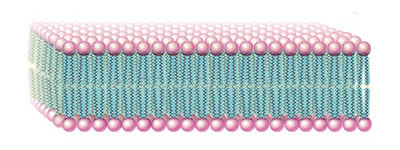
of a typical lipid bilayer