MOLECULAR & CELLULAR
NEUROBIOLOGY
Master Course Cognitive Neuroscience - Radboud
University, Nijmegen
|
|
Chapter 5: Molecular biological research methodology |
Bioinformatics - data analysis | CRISPR-cas genome editing |
ChIP-chip/seq |
|
Genetic transmission
All hereditary information is transmitted from one generation to the other through DNA. The basic hereditary unit, a gene, consists of a distinct fragment of DNA which encodes a specific polypeptide (protein). Each individual has two copies of each gene, called alleles, one from the mother and one from the father. The ~21,000 genes of the human genome are localized in a linear sequence along 23 pairs of chromosomes, including 22 pairs of autosomes (chromosomes 1 to 22) and one pair of sex chromosomes, X and Y. Females have two X chromosomes, while males carry one X and one Y chromosome. Each parent must contribute one of each chromosomal pair and thus one copy of each gene. The gene is located at a particular site on the chromosome and is referred to as the chromosomal locus or genetic locus. A given gene always resides at the same genetic locus on a particular chromosome so the loci on homologous chromosomes are identical. However, alleles residing at these loci may be homozygous (identical alleles) or heterozygous (two different alleles). Classification of inherited disorders The DNA molecule is notable for its stability and seldom changes from one generation to the other. Nevertheless, occasional base sequence changes (mutations) do occur . Mutations are defined as stable sequence changes in DNA that are inherited. Mutations occur at a frequency of approximately one every 200 years. Mutations may involve a portion of the chromosome, a single nucleotide as either a substitution, a deletion, or an insertion, or multiple nucleotides. It is thus convenient to classify heredity diseases into three broad categories, as follows: (1) chromosomal abnormalities; (2) single-gene or monogenic disorders; (3) polygenic disorders or complex traits which are due to interactions of multiple genes and nongenetic factors. Chromosomal abnormalities Human cells each have two copies of each chromosome (diploids) and each chromosome has two arms referred to as the long (Q) or the short (P) arms (Fig 1). The arms of the chromosome meet at primary constriction referred to as the centromere. Chromosomal abnormalities are common and are the most common cause for spontaneous abortions. Chromosomal abnormalities are much more a concern of pediatrics than of adult disease. The chromosomal abnormalities are usually large and can be detected most of the time by doing karyotyping or simply microscopic analysis of the chromosomes. They will not be discussed except to state that the two most common adult cardiovascular chromosomal diseases are Down syndrome (Trisomy 21) and Turner syndrome (xo), both due to non-disjunctions of the chromosomes. Non-disjunction refers to the failure of a homologous pair of chromosomes to separate during meiosis. When an additional copy of a chromosome is added during fertilization, three copies of the same chromosome (Down syndrome) or only one copy (Turner syndrome) is found in the zygote rather than a chromosome pair. |
|
Single-gene disorders 
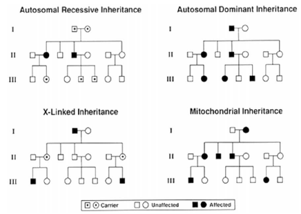
A single-gene disorder is an inherited disease caused by mutations in a single gene that are necessary and sufficient for the development of the phenotype. They show a Mendelian pattern of inheritance classified as autosomal-dominant, autosomal-recessive, or X-linked (dominant or recessive). Mitochondria has its own DNA which encodes for 37 genes. Diseases due to mitochondrial DNA mutations are only transmitted from the mother (no male-to-male transmission), since only ovum has mitochondria. It is important to realize that only a very small fraction of cardiovascular disorders are single-gene disorders and in the whole population the prevalence of single-gene disorders is rare, varying from 1 in 1000 to 1 in 10,000 and essentially never exceeds 1 in 500 (0.5%). It is estimated there are about 14,000 single-gene disorders, of which over 1500 of the genes have been identified. The genotype refers to the genetic basis, while phenotype refers to the observable features such as height, weight, or clinical features of a disease. One may possess the gene but not express the phenotype. The percentage of individuals with a gene that is expressed as a phenotype is referred to as penetrance and the variability in the clinical features of a particular expressed phenotype is termed expressivity. On average, a mutation occurs every 106 cell divisions or once every 200,000 years. Only mutations occurring in the gametes are transmitted. The patterns of inheritance are shown in the diagram in Fig 2. In autosomal-dominant disorders males and females are equally affected; an offspring of an affected parent will have a 50% chance of inheriting the mutant allele. In sporadic cases, the mutations occur de novo in one of the germ lines of the parents but by definition is absent in the somatic cells of parents. Autosomal-dominant inheritance usually has variable expressivity. The following features are characteristic of autosomal-dominant inheritances (Fig 2): (1) each affected individual has an affected parent unless the disease occurs due to a new mutation or there is low penetrance; (2) there is usually an equal split (50/50) of normal and affected offspring born to an affected individual; (3) normal children of an affected individual will have only normal offspring; (4) equal portions of males and females are affected; (5) both sexes are equally likely to transmit the abnormal allele to male and female offspring and male-to-male transmission occurs; and (6) vertical transmissions through successive generations occur. Two other characteristic features that help differentiate this type of inheritance for autosomal-recessive disorders are delayed age of onset and variable clinical expression. In autosomal-dominant, the phenotype is observed despite only one of the gene or alleles being affected. In autosomal-recessive inheritance, both alleles are affected, otherwise there is no phenotype. Males and females are equally affected. Clinical uniformity is more typical and disease onset generally occurs much earlier in life than in autosomal-dominant. Recessive disorders are more commonly diagnosed in childhood and, on average, only one in four children or 25% will be affected. The following are characteristic of autosomal-recessive disorders: (1) parents are clinically normal in alternate generations (genetically are heterozygous); (2) alternate generations are affected, with no vertical transmission; (3) both sexes are affected with equal frequency; (4) each offspring of heterozygous carriers has a 25% chance of being affected, a 50% chance of being an unaffected carrier, and a 25% chance of inheriting only normal alleles. |
|
FIG 2. Pedigrees outlining the usual inheritance patterns for autosomal-dominant and recessive traits, X-linked inheritance, and mitochondrial inheritance. Squares signify males and circles signify females. Filled circles and squares are affected females and males, respectively. |
X-linked inherited disorders are caused by defects in genes located on the X chromosome. Females have two X chromosomes and thus, if only one mutant allele, may seldom develop the phenotype. On the other hand, males have a single X chromosome and are more likely to display the full syndrome whenever an abnormal gene is inherited from the mother. The characteristic features of X-linked inheritance include (1) no male-to-male transmission; (2) all daughters of affected males are carriers; (3) sons of carrier females have a 50% risk of being affected and daughters have a 50% chance of being carriers; (4) affected homozygous females occur only when an affected male and carrier female have children; and (5) the pedigree pattern in X-linked recessive traits tends to be oblique because of the occurrence of the trait of the sons of normal carriers but not in sisters of affected males. Examples of X-linked disorders of the heart include X-linked cardiomyopathy, Bart syndrome, a Duchenne/Becker, and Emery–Dreifuss muscular dystrophy. Since mitochondrial DNA is transmitted to the next generation only by the female, DNA mutations can only be inherited by the mother. The characteristic features of mitochondria inheritance include the following: (1) equal frequency and severity of disease for each sex; (2) transmission through females only, with offspring of affected males being unaffected; (3) all offspring of affected females may be affected; (4) extreme variability of expression of disease within a family; (5) phenotype might be age-dependent; (6) organ mosaicism is common.
Identification of genes causing polygenic disorders - the new frontier
|
|
Next page: Genetic mapping | Go back to: Techniques used in Molecular Biology |
|