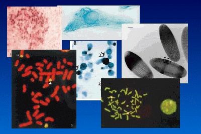
In situ hybridization
In situ hybridization, as the name suggests, is a method of localizing either mRNA within the cytoplasm or DNA within the chromosomes of the nucleus by hybridizing the sequence of interest to a complimentary strand of a nucleotide probe. Normal hybridization requires the isolation of DNA or RNA, separating it on a gel, blotting it onto nitrocellulose and probing it with a complimentary sequence (see under "Detection of DNA, RNA and protein"). The basic principles for in situ hybridization are the same, except one is utilizing the probe to detect specific nucleotide sequences within cells and tissues. The sensitivity of the technique is such that threshold levels of detection are in the region of 10-20 copies of mRNA or DNA per cell. In situ hybridization may present a unique set of problems as the sequence to be detected could be at a low concentration, be masked because of associated protein, or protected within a cell or cellular structure. Therefore, in order to probe the tissue or cells of interest one has to increase the permeability of the cell and the visibility of the nucleotide sequence to the probe without destroying the structural integrity of the cell or tissue. We also have to consider the type of probe to use, and how best to label it, to give the best level of resolution with the highest level of stringency. Some of these choices are dependent on the type of questions being asked, facilities available, and how one intends to assess the outcome, i.e. qualitatively or quantitatively. There are almost as many methods for carrying out in situ hybridization as there are tissues that have been probed. So more important than having a recipe is to have an understanding of the different stages in the process and their purpose.
Choise of probes. Probes are complimentary sequences of nucleotide bases to the specific RNA or DNA sequence of interested. There are essentially four types of probe used:
1. Oligonucleotide probes. These are produced synthetically by an automated chemical synthesis.
2. Single-stranded DNA probes. These have similar advantages to the oligonucleotide probes except they are much larger, probably in the 200-500 bp size range.
3. Double-stranded DNA probes
4. RNA probes or Riboprobes. RNA probes have the advantage that RNA-RNA hybrids are very thermostable and are resistant to digestion by RNases.
Choice of labelling probes. Traditionally, probes were radiolabelled with 32P 35S of 3H but more recently non-radioactive labels have been developed which in general are more versatile, have equal or greater sensitivity, produce quicker results generally, and also of importance, avoids the handling of hazardous materials. Radiolabelled probes are however still the choice for many workers. 32P and 35S are the most commonly used radioisotopes because their high activity is necessary to detect transcripts present in low amounts. In spite of this, incubation times frequently span several weeks. Radiolabelled probes are of course visualized by exposure to photographic emulsions which are then developed. In in situ hybridization, instead of using photographic film, the slide containing the section of interest is dipped into a photographic emulsion which is allowed to dry. The slide is stored in the dark at -80oC to allow the slide emulsion to become exposed. After the incubation period, the slides are then eveloped, as would be a normal photographic film. Thus, the section or cells of interest are viewed through the developed photographic emulsion and the black silver grains indicate the sites of the labelled transcripts.
There are an increasing number of non-radioactive labels which are available with which to label probes. Some such as biotin, and digoxigenin require an additional step for colour development other labels such as fluorescein and rhodamine can be linked directly to the nucleotide.
Controls. The most important part of any experimental procedure is the inclusion of controls. In carrying out in situ hybridization one has to be confident that the hybridization reaction is specific and that the target sequence is in fact being labelled and not other components of the cell. In addition, if no staining is observed, is there a problem with tissue preparation? Therefore, one also needs controls to make sure that sample preparation was good.
Types of in situ hybridization:
- in situ hybridization of frozen sections
- whole-mount in situ hybridization
- in situ hybridization on cultured cells