MOLECULAR & CELLULAR NEUROBIOLOGY 
Master Course Cognitive Neuroscience - Radboud University, Nijmegen


INDEX

INTRODUCTION CELLS AND WITHIN CELLS IN A NUTSHELL GENOMICS MOLECULAR BIOLOGICAL RESEARCH METHODOLOGY NEURODEVELOPMENT  


Chapter 5: Molecular biological research methodology

           Molecular biology and Recombinant DNA technology Detection of DNA, RNA and protein Generation of gene expression atlases of the CNS
           Techniques used in Molecular Biology    Detection of RNA Gene transfer - transgenic animals
           Genetic transmission    In situ hybridization Optogenetics
           Genetic mapping    PCR Cloning
           Genomic and cDNA libraries    Microarray and RNA-seq analysis Stem cells
   Bioinformatics - data analysis    CRISPR-cas genome editing
  ChIP-chip/seq  


Optogenetics                                                                                                              

Understanding how different kinds of neurons in the brain work together to implement sensations, feelings, thoughts, and movements, and how deficits in specific kinds of neuron result in brain diseases, has long been a priority in basic and clinical neuroscience. Optogenetic tools are genetically encoded molecules that, when targeted to specific neurons in the brain, enable their activity to be driven or silenced by light and they have made a significant impact on neuroscience, enabling specific modulation of selected cells within complex neural tissues. These molecules are microbial opsins (seven-transmembrane proteins) which react to light by transporting specific ions across the lipid membranes of cells in which they are genetically expressed. These tools enable the causal assessment of the roles that different sets of neurons play within neural circuits, and are accordingly being used to reveal how different sets of neurons contribute to the emergent computational and behavioral functions of the brain. In general, optogenetics, as the term has come to be commonly used, thus refers to the integration of optics and genetics to achieve gain or loss-of-function of well-defined events within specific cells of living tissue. Optogenetic tools are also being explored as components of prototype neural control prosthetics capable of correcting neural circuit computations that have gone awry in brain disorders.

Opsins (light-driven ion pumps) had been studied since the 1970s because of their fascinating biophysical properties—these molecules undergo fast and rich conformational changes during light-driven ion transport—and also because of the ecological and organismal insights they reveal into how different life forms use light as an energy source or sensory cue (Figures 1 and 2). Bacteriorhodopsin, for example, discovered in the early 1970s in the archaeon Halobacterium salinarum, pumps protons out of cells in response to green light (Figure 1A and 2A). In the late 1970s, the related molecule halorhodopsin, an orange light-driven inward chloride pump (Figure 1B and 2A), was discovered in the same organism, which lives in high salinity environments where these two ion transport rhodopsins contribute to the bioenergetics of the organism by pumping protons out of, and chloride into, cells in response to light. The opsin that drives phototaxis in the green alga Chlamydomonas reinhardtii was a light-gated cation channel that, when illuminated, lets positively charged ions (such as H+ and Na+) pass into cells in which it is heterologously expressed and accordingly the molecule was named channelrhodopsin-2 (Figure 1C and 2A). It can be used to depolarize cells such as oocytes or HEK (Human Embyonic Kidney) cells in response to light, and therefore channelrhodopsin-2 is a useful tool to manipulate intracellular Ca2+ concentration or membrane potential, especially in mammalian cells. Using a light-activated channel requires, for in vivo use, an implanted optical fiber to be inserted into the brain. This light-driven method found rapid adoption by the neuroscience community.


Figure 1. Adaptation of microbial opsins from nature for the optical control of neural activity. Diagrams depicting the physiological responses of (A) archaerhodopsins and bacteriorhodopsins (light-driven outward proton pumps), (B) halorhodopsins (light-driven inward chloride pumps), and (C) channelrhodopsins (light-gated inward nonspecific cation channels), when expressed in the plasma membranes of neurons and exposed to light.


Figure 2. Basic properties of known single-component optogenetic tools with spectral and kinetic information.

(A) Single-component optogenetic tool families; transported ions and signaling pathways are indicated. Channelrhodopsins (ChR) conduct cations and depolarize neurons upon illumination (left). Halorhodopsins (HR) conduct chloride ions into the cytoplasm upon yellow light illumination (center, left). Bacteriorhodopsins (BR) are light-driven outward proton pumps (center, right; PR is retinal protein phoborhodopsin, also called sensory rhodopsin II). OptoXRs are rhodopsin-GPCR (G protein–coupled receptor) chimeras that respond to green (500 nm) light with activation of the biological functions dictated by the intracellular loops used in the hybrid (right).

(B) Kinetic and spectral attributes of optogenetic tool variants for which both of these properties have been reported and for which minimal activity in the dark is observed. Visible spectrum shown; not venturing into the ultraviolet is preferred, for safety and light penetration reasons, although the 450–470 nm peak probes also can be excited very effectively with UV light (~360–390 nm). Decay kinetics are plotted against peak activation wavelength only to demonstrate groupings and classes over the range of spectral and temporal characteristics and the feasibility of dual channel control using tools that are well separated in the spectral and temporal domains.


Delivering optogenetic tools into neuronal systems

A successful neuroscience experimental paradigm requires specific in vivo targeting of the optogenetic tool (Figures 3 and 4). Major categories of in vivo delivery and targeting strategies include (1) viral promoter targeting, (2) projection targeting, (3) transgenic animal targeting, and (4) spatiotemporal targeting—subsets of which may be combined for further increased specificity.

(1) Viral expression systems have numerous advantages for optogenetics, including rapidity and flexibility of experimental implementation, potency linked to high gene copy number, and capability for multiplexing genetic and anatomical specificity. Indeed, viral vectors currently represent the most popular means of delivering optogenetic tools to intact systems. Viral expression systems thus have the dual advantages of fast/versatile implementation and high infectivity/copy number for robust expression levels. Cellular specificity can be obtained with viruses by specific promoters (if small, specific, and strong enough), by spatial targeting of virus injection, and by restriction of opsin activation to particular cells (or projections of specific cells) via targeted light delivery. Lenti (LV) and adeno-associated (AAV) viral vectors have been used successfully to introduce opsins into the mouse, rat, and primate brain. Additionally, these have been well tolerated and highly expressed over long periods of time with no reported adverse effects. LV may be easily produced using standard tissue culture techniques, while AAV may be more challenging to produce within standard laboratory environments and can be produced either by individual laboratories or through core virus production facilities. AAV-based expression vectors display low immunogenicity and offer the advantage of viral titers that result in larger transduced tissue volumes compared with LV. Additionally, AAV is considered safer than LV since currently available strains do not broadly integrate into the host genome. The high multiplicity-of-infection achieved with LV and AAV is particularly useful for optogenetics, as high copy numbers of opsin genes are required to ensure robust photocurrent responses in vivo. LV is more restricted in its diffusion in vivo than AAV and can be used to target subfields of a structure such as the CA1 region of the mouse hippocampus. A major downside of viral expression systems is a maximum genetic payload length; only promoter fragments that are small (less than 4 kb), specific, and strong may be used, and these are rare. Several potential different recombinase-dependent viral vector designs have emerged and a Cre recombinase-dependent double-floxed inverted opsin gene in AAV under the EF1a promoter was ultimately found to provide a suitable combination of strength and specificity to enable behaviorally significant optogenetic gain or loss of function within the constraints of the freely moving mammal system. Not only is this strategy versatile in the sense that it can be applied at will to the large and growing pool of Cre driver lines, soon to include rat as well as mouse lines, but this approach is also by design expandable along new dimensions that enable combinatorial experiments (Figure 3).
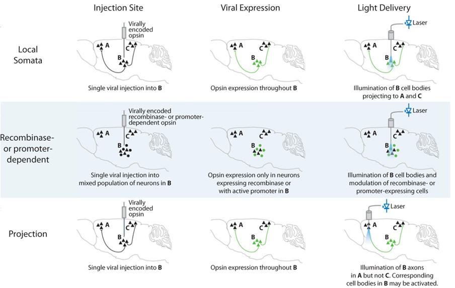
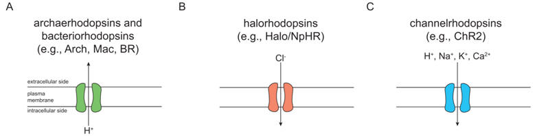
(2) Projection targeting. Cells could look the same from the genetic standpoint but serve fundamentally different functions by virtue of differential wiring. Projection targeting is the ability to selectively drive or inhibit cells defined by their wiring or projections. Microbial opsin gene products, especially with assistance from molecular engineering such as the addition of cellular trafficking motifs may traffic down dendrites or axons and create light-sensitive projections. This property, in the setting of anatomical specificity provided by viruses, allows transduction of cell bodies in one brain region and illumination of axonal projections in another (Figure 3C), thereby defining a cell population for excitation or inhibition by virtue of its connectivity. This approach provides a versatile promoter-independent means to control cells, requiring only anatomical information, and even with simple light guidance strategies this method can be applied to projections as short as hundreds of micrometers. A caveat of this approach is that all local photosensitive axons will be driven by light, even fibers of passage that do not synapse in the illuminated region.

(3) Delivering optogenetic tools into animals. Generation of mouse transgenic lines directly expressing opsin genes under local promoter-enhancer regions provides a distinctly useful means of achieving cell-type-specific opsin expression. While transgenic mouse lines require effort, time, and cost associated with their production, validation, and maintenance, the convenience and reliability of homogeneous opsin-expressing animals provides major experimental leverage. The use of transgenic or knock-in animals obviates viral payload limitations and thus allows for tighter control of transgene expression using larger promoter fragments or indeed the endogenous genome in full via knock-in.

(4) Spatiotemporal targeting. Cells may also be targeted by virtue of their birthdate or proliferation status, location at a moment in time, and other versions of what might be called spatiotemporal targeting; this approach has reached its most advanced state in the course of targeting specific neocortical layers. A long-sought goal of neuroscience has been to tease apart the role of specific layers, and of layer-specific neurons, in cortical microcircuit processing, brain-wide network dynamics, and animal behavior. In utero electroporation (IUE) may be employed to target opsins to distinct layers of the cortex, capitalizing on the sequential layer-by-layer ontogeny of neocortex in mammals, by incorporating the DNA into neurons generated during a specific embryonic stage. Beyond this special targeting capability, an additional unique advantage of IUE is that opsins are expressed from before the time of litter birth (allowing electrophysiological experiments at a younger stage than with viral expression). In principle, cells may also be targeted for optogenetic control by (i) active proliferation status at a particular moment in time, using cellcycle-dependent Moloney-type retroviruses; (ii) location at a particular moment in time (e.g., via migration through a particular anatomical location during development; and (iii) other methods including ex vivo sorting followed by transduction and transplantation. In general, the range of genetic techniques for delivering opsin genes into the brain has become broad and versatile.


Figure 3. Targeting optogenetic tools in vivo.

Upper: Direct stimulation of neuronal cell bodies is achieved by injecting virus at the target region and then implanting a light-delivery device above the injected region. Even this simple experiment can provide specificity with viruses that will not transduce afferent axons and fibers of passage.

Middle: Additional cell-type specificity is attained either by cell-type-specific promoters in the viral vector or via a recombinase-dependent virus, injected in a transgenic animal expressing a recombinase such as Cre in specific cells, leading to specific expression of the transgene only in defined cell types.

Bottom: Projection (axonal) targeting is achieved by viral injection at the region harboring cell bodies, followed by implantation of a light-delivery device above the target region containing neuronal processes from the virally transduced region; in this way cell types are targeted by virtue of their projections.


.


Figure 4. Spatially localized stimulation of opsin-expressing cells. Direct light activation of an opsin-expressing neuron is restricted only to the genetically targeted cells. While neurons not expressing an opsin fail to be activated by the same light pulse, opsin expressing neurons rapidly depolarize leading to action potential generation  


Examples of results


Figure 5. Use of opsins to mediate light-driven neural silencing or light-driven spiking.

Left: Demonstration of use of Halobacterium sodomense archaerhodopsin-3 (Arch) to mediate light-driven neural silencing in cortical pyramidal neurons of awake mice. Top: Neural activity in a representative neuron before, during, and after 5 seconds of yellow light illumination, shown as a spike raster plot (upper panel), and as a histogram of instantaneous firing rate averaged across trials (lower panel; bin size, 20 ms). Bottom: Population average of instantaneous firing rate before, during, and after yellow light illumination (black line, mean; gray lines, mean ± SE).

Middle: Demonstration of use of Natronomonas pharaonis halorhodopsin (Halo/NpHR) to mediate light-driven spike quieting, demonstrated for a representative hippocampal neuron in vitro. Top: (“Current injection”), neuronal firing of 20 spikes at 5 Hz, induced by pulsed somatic current injection (~300 pA, 4 ms). Middle: (“Light”), membrane hyperpolarization induced by two periods of yellow light, timed so as to be capable of blocking spikes 7–11 and spike 17 out of the train of 20 spikes. Bottom: (“Current injection + Light”), yellow light drives Halo to block neuron spiking (note absence of spikes 7–11 and of spike 17), while leaving intact the spikes elicited during periods of darkness.

Right: Demonstration of use of Chlamydomonas reinhardtii channelrhodopsin-2 (ChR2) to mediate light-driven spiking in two different hippocampal neurons, in response to the same train of blue light pulses (with timings selected from a Poisson distribution with mean interval λ = 100 ms).


Figure 6. An example of behaviour probing using optogenetics. The nucleus accumbens is probed specifically; applying both localized expression of the desired opsin as well as correct placement of the optical fibre. Using the appropriate light wavelength, a specific cell population can be activated (green cells), resulting in their selective activation within intact neural network. This setup enables investigation of the functional role of distinct brain areas, neural network, or cell type in behaviour of freely moving animals.  


Future of optogenetics

The optogenetic toolbox has broadly expanded to include proteins that are powerful and diverse in their ionic selectivity, spectral sensitivity, and temporal resolution. Combined with powerful molecular techniques for transgenic and viral expression in rodents, zebrafish, and flies, the current generation of optogenetic tools may be adapted to many questions within Neuroscience. The current generation of optogenetic tools has been optimized for stronger expression, higher currents, and spectral shifts to allow combinatorial control within the same volume of space. Ongoing improvements to the toolbox will yield molecular tools targeted to subcellular compartments (such as dendrites or axons), tools for two-photon activation, and tools that further expand the optical control of biochemistry.


Next page: Cloning Go back to: Gene transfer - transgenic animals