|
MOLECULAR & CELLULAR
NEUROBIOLOGY
Master Course Cognitive Neuroscience - Radboud
University, Nijmegen
|
|
|
|
Chapter 5: Molecular biological research methodology |
| Bioinformatics - data analysis | CRISPR-cas genome editing |
| ChIP-chip/seq |
|
|
Generation of gene expression atlases of the CNS
An understanding of the molecular mechanisms of development, function and dysfunction in the mammalian CNS will ultimately require both precise knowledge of the distribution of cell types expressing molecules of interest, and the ability to explore mechanisms at the level of single cells or cell types. The brain is the most complex organ in our body, responsible for perception, behavior, cognition, memory, and consciousness. It is comprised of about a trillion nerve cells, or neurons, whose intricate and precisely wired connections underlie all of these functions. But the neurons are not all the same: many thousands of different classes of neurons, defined by a variety of criteria such as morphology, patterns of connectivity, and expression of particular neurotransmitters and receptors, serve as the cellular building blocks of the brain. Each of these neuronal types has a specific physiological role in brain function. Neurological and psychiatric diseases are diseases of particular neurons or neural circuits.
Using histological tools like the Golgi staining method, neuroanatomists in the late 19th and early 20th centuries defined hundreds of different neuronal cell types based on their location in the brain, morphology, and connectivity. Throughout the 20th century, this number grew, and neuroscientists were also able to assign particular physiological functions to a large number of these neuronal types and many of the circuits linking these cells. At present, the number of distinct neuronal cell types is not precisely known, but it is estimated that there must be at least several thousand. This estimate is supported by the finding of novel subclasses whenever a particular population of neurons is studied in detail. These thousands of cell types, through their specific patterns of interconnections, direct the functioning of the brain.
Subpopulations of neurons are increasingly being distinguished based on their patterns of gene expression. Historically, molecular neuroanatomy started with cellular pharmacology, the definition of the neurotransmitters and neurotransmitter receptors made by particular neurons, using a variety of physiological, in situ radioligand binding, and histochemical methods. The development of the technique of immunohistochemistry accelerated the mapping of proteins (such as neuropeptides) and other epitopes (such as specific carbohydrate moieties) to particular neurons in cases where a suitable antibody was available. However, it is the development of sensitive and reproducible mRNA in situ hybridization techniques that unleashed the systematic analysis of gene expression in neurons, because this approach can be readily applied to all genes without requiring the labor-intensive development of specific detection reagents (such as antibodies); in all cases, a small gene fragment is sufficient to develop an appropriate in situ hybridization probe. In situ hybridization methods have been supplemented by transgenic (promoter-based, and bacterial artificial chromosome-BAC-based) and knock-in approaches, which make it possible to visualize the pattern of expression of particular genes using genetically encoded reporters driven from the gene locus in transgenic mouse lines.
The complexity of brain cell types and circuits is reflected in the complexity of gene expression patterns in the brain. It is believed that perhaps a third to half of all genes are largely or exclusively dedicated to directing the development, maintenance and functioning of the brain. With about 30,000 genes in the human genome, the task of mapping all genes to the many thousands of neuronal classes and neuronal circuits (molecular neuroanatomy) might seem beyond reach. In fact, this mapping is taking place. It is proving to be remarkably informative and such studies have shown that analysis of gene expression in neurons can yield essential information on neural development and function, such as the identity of neurons involved in responses to particular drugs, or the genes that control the development of particular classes of neurons. Such analysis has also defined molecular markers of particular neuronal cell types, helping with the taxonomy of brain cell types and the division of known neuronal classes into further subclasses. The availability of cell type-specific molecular markers for particular neuronal classes has provided tools to deliver genes and gene products to those neurons, facilitating the analysis of their development, connectivity, function, and dysfunction. The identification of genes expressed in particular classes of neurons linked to specific diseases provides new drug targets for the treatment of a wide range of ailments including stroke, spinal cord injury, neurodegenerative diseases like Parkinson’s disease, brain tumors, schizophrenia, depression, anxiety disorders, and addiction. Neuronal cell type-specific markers provide a means for developing gene therapies that involved changing gene expression in particular neurons. They also make it possible to visualize neural circuits in their normal and abnormal states, which is likely to have a large impact on the diagnosis of disease and the evaluation of the effectiveness of therapy. The identification of transcription factors that control cell fate and connectivity in the brain will accelerate the development of therapies to regenerate nervous tissue.
In the 1990s, the Brain Molecular Anatomy Project (BMAP) was launched, a series of funding initiatives to map gene and gene product expression to neuronal cell types to create a Molecular Brain Map (see figure below). The GENSAT project aims to systematically map the expression patterns of thousands of genes in histological sections of the mouse brain and spinal cord in the adult and at three stages of development (embryonic day E10.5, E15.5, and postnatal day P7), including map gene expression through high-throughput in situ hybridisation and a large-scale screen using transgenic mice carrying modified BACs containing an enhanced green fluorescent protein (EGFP) reporter gene has been performed to map gene expression at the cellular level. A library of verified BAC vectors and a series of transgenic mouse lines have been identified that offer genetic access to CNS regions, cell classes, and pathways. Novel insights into the functions of individual genes, into principal steps in the development of the CNS (e.g. cell specification, migration, axon targeting) and into the organization, function and dysfunction of the mature brain have emerged from these studies. The anatomical data and library of BAC vectors generated in this screen provide a rich resource for discovery of molecular mechanisms impacting CNS development and function, and enable a broad array of investigations not previously available to the neuroscience community.
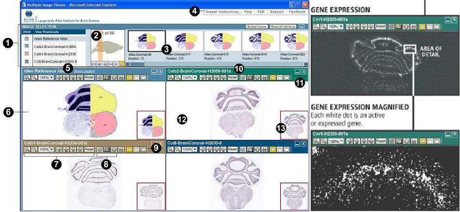
(numbers in the figure indicate the various possibilities in the brain atlas tour).
- - A dataset of full-length transcripts/cDNAs expressed in the nervous system; this dataset is well on its way to completion.
- - A bank of Bacterial Artificial Chromosomes (BACs) that permit transgenic labeling and manipulation of major neuronal populations in the mouse brain; only a few hundred of these exist today.
- - A bank of short promoter elements for extending such manipulations to primates and other non-genetic systems; few of these exist at present.
- - A set of antibody probes (for immunohistochemistry) that help extend and leverage the Molecular Brain Map. A priority is the generation of antibodies to the ~1,500 transcription factors encoded in the genome, to help identify neuronal cell types and stem cells; only a small fraction (~10%) of these exist at present.
- - Improved methods for isolation of high-quality mRNA from small numbers of neurons, including laser-capturing of mRNA from neurons identified through expression of a transgenic reporter (such as GFP), and for faithful linear amplification of cDNA from the mRNA for gene expression analysis.
- - Improved methods for mapping neuronal circuits (identifying all inputs to each neuron and all outputs from each neuron)
- - Methods for detecting electrical activity in mammalian neurons by optical recording using genetically-encoded reporters
- - Methods for controlling activity in defined neurons, in particular genetically-encoded modulators of electrical activity that can be activated with specific signals such as pharmacological agents or light
- - Methods for detecting neuronal activity in deep brain structures using such genetically-encoded reporters
- - Methods for mapping patterns of neuronal activity onto patterns of gene expression and neuronal interconnectivity
- - Methods for persistent labeling of neurons over time, to follow plastic changes in morphology, connections, or function
- - Methods for visualizing changes in neuronal connections, such as pulse-chase labeling of synapses.