 Some recent
experiments have revealed interesting actions of protein kinases on GABAa receptors.
Some recent
experiments have revealed interesting actions of protein kinases on GABAa receptors.Some recent
experiments have revealed interesting actions of protein kinases on GABAa receptors.
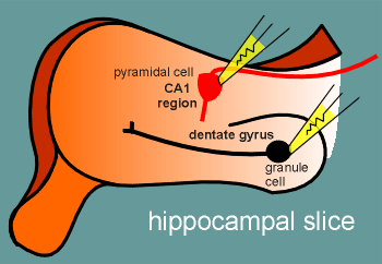
The experiments were conducted with thin slices of brain tissue bathed in a physiological incubation medium (this is a common method to study neurons in vitro).
The experiments in question were conducted on slices from a particular area of the brain called the hippocampus (more on the hippocampus).
The experiments involved patching two different neuronal cell types in the tissue, the so-called pyramidal cells (in an area of the hippocampus called the CA1 region) and granule cells (in a region of the hippocampus called the dentate gyrus).
The cells were patch in the whole cell configuration (review patch-clamp configurations?).
Thus molecules (such as active protein kinases) could be introduced into the cells via the patch electrode.
The experiments examined the action of protein kinase A (PKA) and protein kinase C (PKC) on the functioning of the GABAa receptor (review production and function of PKA and PKC?).
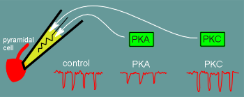 The first experiments were conducted with the pyramidal cells.
In control experiments miniature inhibitory postsynaptic potentials (mIPSP) were recorded.
These are brought about by spontaneous release of presynaptic GABA.
Typical mIPSP in a control experiment are illustrated above.
In another set of experiments active PKA was in the patch electrode and thus, when the electrode went into the cell attached configuration, was introduced into the cell (the researchers gave the kinase a certain pretreatment to ensure that it was in an active form).
In these experiments the mIPSP were clearly smaller (results of typical experiment shown above).
Finally, in a third set of experiments active PKC was introduced into the cell.
The PKC had no clear effect on the size of the mIPSP and the researchers concluded that the GABAa receptors of the pyramidal cells of the CA1 region are affected only by PKA.
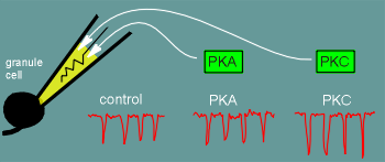 When the granule cells of the dentate gyrus were studied the results were
very different.
When the granule cells of the dentate gyrus were studied the results were
very different.
Here PKA had no clear effect and PKC potentated the mIPSP!
The researchers tentatively conclude that "specific subunits in synaptic GABAa receptors may be responsible for region specific differences".
In other words, the GABAa receptors of the pyramidal cells have a different subunit composition to those of the granule cells such that the former are sensitive to PKA (and give attenuated responses) and the latter are sensitive to PKC (and give potentiated responses).
The researchers conclusions are tentative because it still must be established that the kinase treatments led to a direct phosphorylation of the receptors.