13
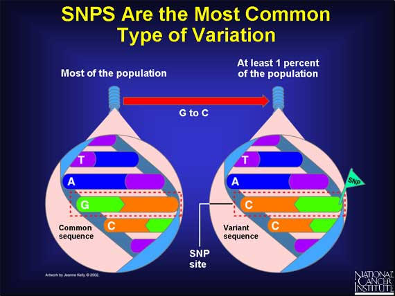
The most common type of genetic variation is the SNP, a single-base change in a DNA sequence that occurs in a significant proportion (more than 1 percent) of a large population. The single base is replaced by any of the other three bases. Here is an example: in the DNA sequence TAGC, a SNP occurs when the G base changes to a C, and the sequence becomes TACC.
14
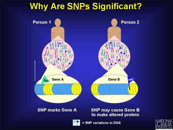
SNPs are scattered throughout the genome and are found in both coding and noncoding regions. SNPs can cause silent, harmless, harmful, or latent effects. They occur with a very high frequency, with estimates ranging from about 1 in 1000 bases to 1 in 100 to 300 bases. This means that there are millions of SNPs in each human genome. The abundance of SNPs and the ease with which they can be measured make these genetic variations significant. Most SNPs occur in noncoding regions and do not alter genes. Scientists are finding that some of these SNPs have a useful function. If a SNP is frequently found close to a particular gene, it acts as a marker for that gene.
The remaining SNPs occur in coding regions. They could alter the protein made by that coding region, which in turn could influence a person's health. To understand how a SNP could do this requires a brief look at proteins and their building blocks, amino acids.
15
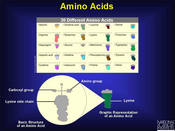
Humans use 20 different amino acids as building blocks to make the thousands of proteins found within the body. Each amino acid has the same basic structure with a central carbon atom that has four groups attached. Three of the groups are the same in every amino acid: one is a single hydrogen, one is an amino group that acts like a hook, and one is a carboxyl group that acts like an eye. The fourth group is a side chain, which is unique for every amino acid.
16
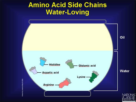
Humans use 20 different amino acids as building blocks to make the thousands of proteins found within the body. Each amino acid has the same basic structure with a central carbon atom that has four groups attached. Three of the groups are the same in every amino acid: one is a single hydrogen, one is an amino group that acts like a hook, and one is a carboxyl group that acts like an eye. The fourth group is a side chain, which is unique for every amino acid. Each side chain has a unique size and shape: some side chains are round and bulky, some are short and skinny, and some are longer than others. Unlike the carboxyl and amino groups, which always like a watery environment, side chains vary in their chemical properties. Some side chains have a charge, and that makes them prefer to be in water.
17
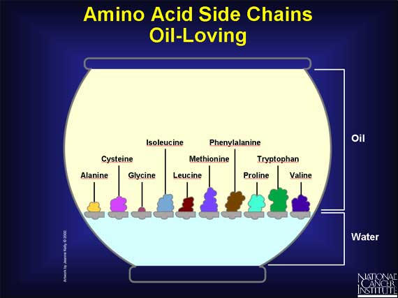
Other side groups have no charge, and that makes them prefer an oily environment. It also prompts them to huddle close together.
18
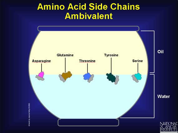
And some side chains are downright indecisive. They usually prefer a watery environment, but tend to adjust themselves to oily ones as well.
19
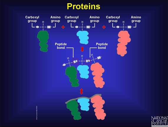
When two amino acids are lined up end-to-end, and the hooks and eyes are allowed to react together, a peptide bond forms. As more amino acids attach, a chain begins to form that has a strong but flexible backbone with side chains sticking out at regular intervals. This is protein-building in progress. But how does the cell do this? How does a sequence of DNA bases turn into a chain of amino acids and form a protein?
20
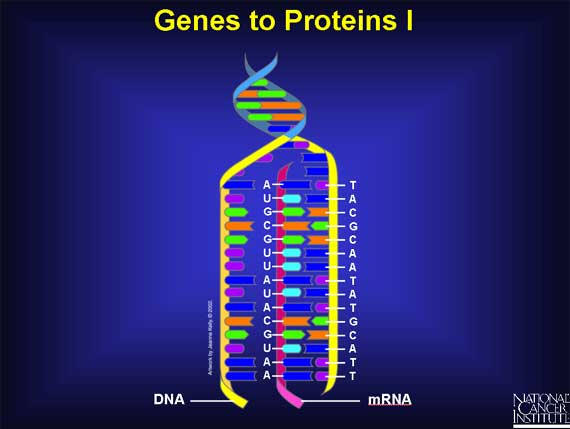
To turn a gene's DNA sequence of bases into a protein's sequence of amino acids requires an intermediary molecule called mRNA. Base pairing is used to create mRNA from one of the DNA strands. Every T in the DNA strand pairs with an A in the mRNA strand, and Cs and Gs in the DNA pair with Gs and Cs in the mRNA. However, if there is an A in the DNA sequence, it pairs with a new base called Uracil (U) in the mRNA strand. Thus, if the DNA sequence is TACGCAATATGCATT, the mRNA sequence becomes AUGCGUUAUACGUAA.
21
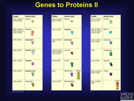
The sequence of the bases of an mRNA (A, U, G, C) directly spells out the sequence of amino acids in the protein. Every three bases in the mRNA sequence codes for a single amino acid and is called a "codon." There are 64 different ways to arrange the four bases in mRNA, e.g., UCA, UCG, AAA, and AGC, and yet there are only 20 different amino acids. This means that one amino acid can have more than one codon. For example, valine has four codons (GUU, GUC, GUA, and GUG), while histidine only has two (CAU and CAC). The mRNA code also has one "start" and three "stop" signals. The codon AUG codes for an amino acid called methionine that always signals the start of protein building. Three other codons --UAG, UGA, and UAA--do not code for any amino acid. Instead, they always signal the mRNA to stop protein building.
22

Proteins are built within a cell structure called a ribosome - a protein factory. First the mRNA attaches itself to the ribosome. Then specific adapter molecules, called transfer RNAs (tRNAs), match themselves to their appropriate codons. Each codon has a corresponding tRNA that carries a specific amino acid at one end. Here is the beginning of a new protein. The first tRNA carrying methionine matched itself to the first codon. Then a second tRNA carrying arginine matched itself with the second codon. A chemical reaction joined one free end (the hook) of the methionine molecule to a free end (the eye) of the arginine molecule, and methionine's tRNA was released. Now a third tRNA carrying tyrosine has matched the third codon, and a chemical reaction has joined the free end of the arginine to tyrosine, and arginine's tRNA has been released. The protein now has three amino acids joined together. This process will continue until a "stop" codon is reached.
23
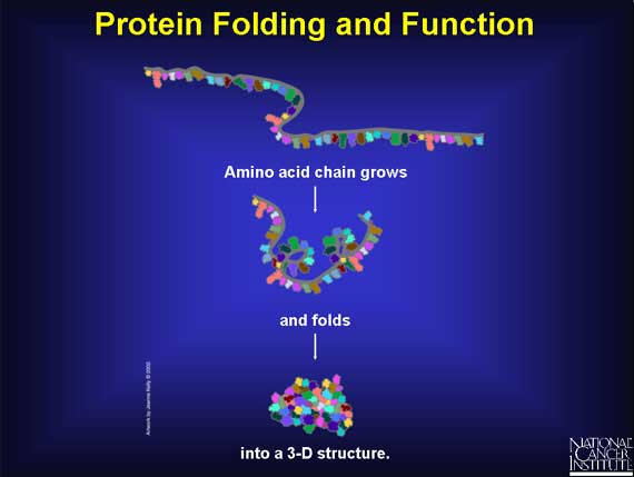
As the amino acid chain grows, it folds into a three-dimensional (3-D) structure, which depends on both the chemical nature and order of the different amino acids. The 3-D structure determines the function of the protein. When there is a change in one or more amino acids, then the ability of the protein to function may be affected. The protein's function may be unchanged or it may become sluggish, hyperactive, or inactive.