MOLECULAR & CELLULAR NEUROBIOLOGY 
Master Course Cognitive Neuroscience - Radboud University, Nijmegen


INDEX

INTRODUCTION CELLS AND WITHIN CELLS IN A NUTSHELL GENOMICS MOLECULAR BIOLOGICAL RESEARCH METHODOLOGY NEURODEVELOPMENT  


Chapter 6: Neurodevelopment

                                                       Neurodevelopment Neuronal outgrowth, axon guidance and axon guidance factors


Neuronal outgrowth and axon guidance


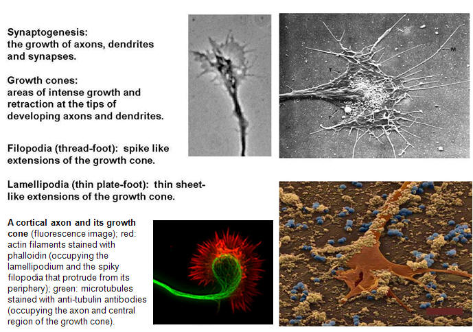
Perhaps the most remarkable feature of the nervous system is its highly ordered connections. We will here consider the processes that insure specific synaptic connections between neurons. Thus, one of the most intriguing problems in developmental neurobiology is that of how growing axons find the correct way to their proper target cells. Often axonal connections are organized in topographic maps, where neighboring cells of the projecting area are connected to neighboring cells in the target area, thus allowing a faithful transfer of positionally stored information from one area to another. A complex "wiring system" of nerve fibers connects the brain's more than 100 billion neurons. Studies on worms, flies, frogs and other animals are now identifying molecules that guide these fibers through the brain to their targets. These discoveries are revealing how the brain develops and may lead to ways of regenerating nerve fibers after injury or preventing wiring defects that result in disease. The brain's wiring system -- nerve fibers (axons) that grow along specific paths to connect with other fibers (dendrites) -- was first described more than a hundred years ago. Scientists learned that axons grow by following an elongated tip called a growth cone (Figure 1). Yet the systems that guide axons to connect with their targets are so complex that they are only now being unraveled. The correct formation of a network by neuronal cells appears to be established by the pathfinding of extending lamellipodia and filopodia at the growth cone. Filopodia, which initiate and elongate from the growth cone lamellipodia, are thin protrusions with actin bundles at their core (Figure 1). Their extension and retraction are initial events in the steering of a growth cone. Clarifying the molecular mechanisms by which filopodia extend and retract, and the signal pathways that control these events, is an important step in understanding how extracellular cues guide growth cones.

  Figure 1


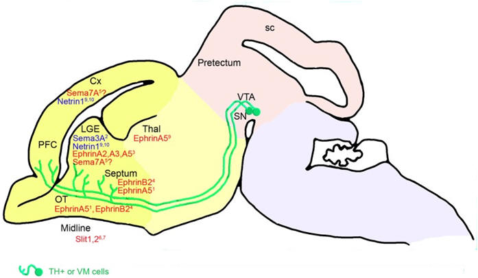
A well-known example often used for neuronal outgrowth/axon guidance studies is the retinal ganglion cell (RGC), the only type of neuron that connects the eye to the brain. The eye is a peripheral outpost of the CNS where the RGCs reside. In the developing vertebrate visual system, retinal ganglion cells in the eye extend axons that navigate over a long distance to their synaptic targets in the midbrain. This impressive navigational feat underlies the precise wiring of the mature brain and is essential for building functional nerve connections. RGC axons navigate to their targets in a remarkably stereotyped and error-free manner and it is this process of directed growth that underlies the complex organization of the adult brain. The RGCs are the only retinal neurons to project into the brain and their peripheral location makes them an unusually accessible population of projection neurons for experiments involving in vivo gene transfer, anatomical tracing, transplantation and in vitro culture. These connections must thus be formed with extreme precision in order for an organism to obtain an accurate representation of the visual field. The retinotectal projection is the classical model system for studying topographic projections. RGC cell bodies and dendrites reside in the retina, while thier axons follow a stereotypic pathway through the diencephalon to innervate their target neurons in the optic tectum. We can manipulate and observe developing RGCs particularly well in tadpoles of the South African claw-toed frog Xenopus laevis or in zebrafish. With these developing visual systems one can study how neurotrophic molecules influence RGC axonogenesis, axonal vs. dendritic arborization, growth cone navigation, target recognition, and synaptogenesis in vivo (Figure 2).


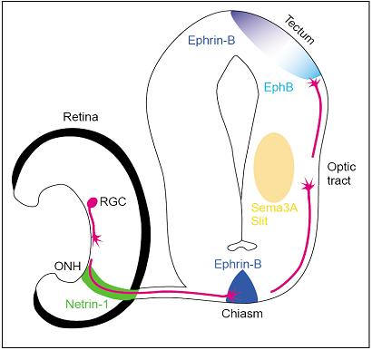
Figure 2. Diagram of the embryonic visual pathway. Guidance molecules belonging to the netrin, slit, semaphorin and ephrin families are expressed in multiple places along the pathway, in discrete segments, and serve to direct the growth of RGC growth cones. For simplicity, the positions of only a few cues are shown. ONH, optic nerve head; RGC, retinal ganglion cell.


The formation of precise neuronal networks is thus critically dependent on the motility of axonal growth cones. Extracellular gradients of guidance cues (see below) evoke localized Ca2+ elevations to attract or repel the growth cone. The polarity of growth cone guidance, with respect to the localization of Ca2+ signals, is presumably determined by Ca2+ release from the endoplasmic reticulum (ER) in the following manner:

(i) Ca2+ signals containing ER Ca2+ release cause growth cone attraction

(ii) Ca2+ signals without ER Ca2+ release cause growth cone repulsion

Recent studies have also shown that exocytic and endocytic membrane trafficking can drive growth cone attraction and repulsion, respectively, downstream of Ca2+ signals. Most likely, these two mechanisms underlie cue-induced axon guidance, in which a localized imbalance between exocytosis and endocytosis dictates bidirectional growth cone steering (see figure below). Thus, polarized membrane trafficking may play an instructive role to spatially localize steering machineries, such as cytoskeletal components and adhesion molecules.


Ca2+ and cyclic nucleotide signaling for growth cone guidance.

(A)   Reception of attractive cue gradient results in the opening of ER Ca2+ channels (RyR and IP3R) on the side of the growth cone facing the cue because ER Ca2+ channels are in the active state due to high ratios of cAMP to cGMP. These Ca2+ signals containing Ca2+-induced Ca2+ release (CICR) or IP3-induced Ca2+ release (IICR) act as an attractive Ca2+ signal.

(B)   Reception of a repulsive cue gradient results only in the opening of plasma membrane Ca2+ channels on the side of the growth cone facing the cue because the ER Ca2+ channels are in the inactive state due to low ratios of cAMP to cGMP. These Ca2+ signals containing neither CICR nor IICR act as a repulsive Ca2+ signal. Black curved arrows: the direction of growth cone turning; red arrows: the direction of Ca2+ mobilization.


Membrane trafficking for growth cone turning.

(A) A gradient of attractive cues evokes attractive Ca2+ signals to facilitate VAMP2-mediated exocytosis on the side of the growth cone facing the cue. The asymmetric insertion of membrane components can provide a driving force for growth cone attraction.

(B) A gradient of repulsive cues evokes repulsive Ca2+ signals to facilitate clathrin-mediated endocytosis on the side of the growth cone facing the cue. The asymmetric removal of membrane components can provide a driving force for growth cone repulsion. Black curved arrows: the direction of growth cone turning; red arrows: the direction of vesicle movement.


Axon guidance factors

We will now deal with the cues that axons use to navigate to their remote targets and accurately map their connections onto these targets, and thus how individual neurons wire themselves together into a precisely interconnected and functional nervous system. We will not go into detail concerning the intracellular signaling mechanisms that translate external signals into specific cellular responses, such as growth cone formation and the elaboration of axonal and dendritic arbors.

To find their proper place in the brain, axons often stretch for several feet, making their way through surrounding tissues and around a myriad of obstacles until they reach their final target. The growth cone then forms a synapse, or a tiny gap where nerve messages are transmitted, with the dendrites of the target neuron. How does this process occur with such remarkable precision? During the 1940s, scientists showed that severed axons from a nerve in a frog's eye always reconnect to the same place in the brain where they were originally attached. These findings suggested that neurons produce chemical labels that identify them and help them recognize their target neurons by a process of  "molecular sensing". About 30 years later, researchers studying animals such as flies, worms and vertebrates finally began to isolate and describe these molecular labels (Figure 3 below). Cell adhesion molecules (CAMs) -- the first to be discovered -- are found on neuron surfaces and bind to similar proteins on nearby cells. By knocking out the genes for specific molecules present in different combinations on different nerve fibers, these proteins were found to help axons recognize and track along paths established by related axons. Growing axons can also change course to follow gradients of certain "attraction" molecules that spread out from target cells and provide long-range cues. Some guidance molecules have other functions. Both short- and long- range "repulsion" molecules were discovered that inhibit axon growth. Some guidance molecules attract certain axons while repelling others. Amazingly, many guidance molecules appear to be remarkably similar in structure and function in different animals -- from fruit flies to humans. An axons response to different molecules is determined by receptors on the surface of the growth cone. When a molecule attaches to these receptors, it causes the growth cone to grow or stop or turn. Cells can change the receptors and other molecules that are active at a given time. Thus, growth cones can respond to different guidance molecules at different stages during their development and change direction. New guidance molecules have been identified and their functions determined (see below). Repulsive guidance molecules may prevent regeneration of axons in the spinal cord and brain after injury, stroke or neurodegenerative diseases. If so, drugs that interfere with these molecules might be developed to make nerve regeneration possible. Knowledge of axon guidance also reveals how the brain develops and may lead to ways of preventing nervous system wiring defects that may underlie disorders such as dyslexia, cerebral palsy and mental retardation.


Figure 3. Classification of factors regulating neurite outgrowth in the vertebrate nervous system (HB-GAM: heparin-binding growth-associated molecule; CSPGs: chondroitin sulfate proteoglycans; NG2: a membrane-spanning chondroitin sulfate proteoglycan; MAG: myelin-associated glycoprotein).


Over the past two decades three experimental approaches have identified a wide variety of novel guidance molecules and their receptors: (1) pairing biochemistry and in vitro tissue culture assays to detect proteins with either attractive or repellent properties; (2) using forward genetics to identify mutations that affect axon trajectories in vivo; or (3) using genetic and tissue culture approaches to characterize the functions of molecules with distributions or molecular structures that make them attractive candidate guidance cues. Using these strategies, four major families of guidance cues (the “canonical cues”) with very well-established roles in neuronal guidance have been identified: the Netrins, Slits, Ephrins, and Semaphorins (see figures below).

Netrins, Slits, and their receptors (for attraction: DCC; repulsion: UNC5 for Netrins; Robo for Slits).


Major A and B classes of Ephrins and their EphA and EphB receptors.


Five classes of vertebrate semaphorins and their receptors(not shown are several non-plexin/neuropilin semaphorin receptors). The key defines distinct molecular domains found in these proteins.


Morphogens and growth factors


While an initial wave of studies in the 1990s was leading to the identification of Netrins, Semaphorins, Ephrins, and Slits as key regulators of axonal attraction and repulsion (see above), parallel studies implicated two other sets of proteins in axon guidance: morphogens of the Wnt, Hedgehog (Hh), and transforming growth factor b (TGFb)/bone morphogenetic protein (BMP) families, as well as a variety of growth factors. Among the morphogens, Wnts have the most widely described axon-guidance functions. Guidance roles for Sonic hedgehog (Shh) in vertebrates have also been described, including being a repellent for a subset of retinal ganglion cells and an attractant for spinal commissural axons. The roles of Hh and TGF-β/BMP proteins in guidance remain to be more fully defined.

A variety of growth factors have also been implicated in attraction of specific populations of axons in the peripheral and central nervous systems of vertebrates. They include hepatocyte growth factor, the neurotrophins brain-derived neurotrophic factor and neurotrophin-3, fibroblast growth factors, glial-derived neurotrophic factor, neuregulin, and stem cell factor. However, the full import of growth factors in axon guidance is poorly understood.


The morphogens Shh and its receptors (Smo, Ptch, Boc, and CDO), and Wnts and their receptors (Frz and Ryk), all of which serve guidance functions; not shown are BMPs and their receptors.


Axon guidance cues serve both neuronal and non-neuronal functions


Repulsive and attractive guidance cues in dopaminergic axon navigation

Understanding how dopaminergic axons navigate through their native environment during development may contribute to increasing efficiency of cell therapy for brain diseases such as Parkinson’s disease. Mesotelencephalic dopaminergic pathways have been studied in great detail because of their implication in major physiological functions as well as in psychiatric, neurological, and neurodegenerative diseases. As an example, the figure on the right shows telencephalic guidance, in particular the expression of repulsive (in red) and attractive (in blue) guidance cues in the environment of mesencephalic dopaminergic (mDA) somas and axons between embryonic day (E) 14.5 and E18.5 in mice.


mDA axons (in green) connect to the telencephalic regions through specific receptors (in green). The cephalic vesicles telencephalon, diencephalon, mesencephalon, and rhombencephalon are delimited in yellow, beige, pink, and purple, respectively. Cx, cortex; LGE, lateral ganglionic eminence; SN, substantia nigra; OT, olfactory tract; PFC, prefrontal cortex; sc, superior colliculus; Thal, thalamus; VTA, ventral tegmental area.


  Go back to: Neurodevelopment