 To do this they used the gene coding for a protein called tau, a
microtuble binding protein present in the axons and terminals of all neurons.
To do this they used the gene coding for a protein called tau, a
microtuble binding protein present in the axons and terminals of all neurons.A beautiful demonstration of the ability of neurotrophins to direct axonal elongation in the developing nervous system has been reported in Nature Neuroscience (Tucker et al. Neurotropins are required for nerve growth during development, Nature Neurosci. 4: 29-37, 2001).
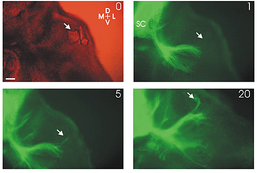
These scientists first developed a transgenic mouse so that growing nerves could be visalized with fluorescent microscopy.
To do this they used the gene coding for a protein called tau, a
microtuble binding protein present in the axons and terminals of all neurons.
They attached to the tau gene the coding sequence for a protein called green fluorescent protein (GFP).
 GFP is the protein responsible for the green fluorescence
displayed by the jelly fish, Aquoria victoria.
GFP is the protein responsible for the green fluorescence
displayed by the jelly fish, Aquoria victoria.
The tau-GFP gene construct was used to generate homozygous animals expressing the construct (the methods used to produce these transgenic animals are beyond the scope of this tutorial, but are similar to those used to produce gene knockout which will explained shortly).
Next, they took tissue slices from the developing nervous system of such animals and introduced beads coated with various neurotrophins and embedded them in the tissue.
Then they followed the growth of the termials over several days.
Shown to the right is a results where a BDNF coated bead (indicated by the white arrow) was implanted and then florescent images taken 1, 5 and 20h later.
Notice how the growing nerve terminals grow towards the BDNF coated beads.
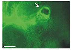 To the left, is a similar experiment with a NGF coated
bead.
To the left, is a similar experiment with a NGF coated
bead.
Here 24h after implantantation the bead is completely covered by florescent nerve terminals.
These kinds of responses could be blocked by introducing antisera to the growth factors, nicely showing that the growth factors are directing the growth.